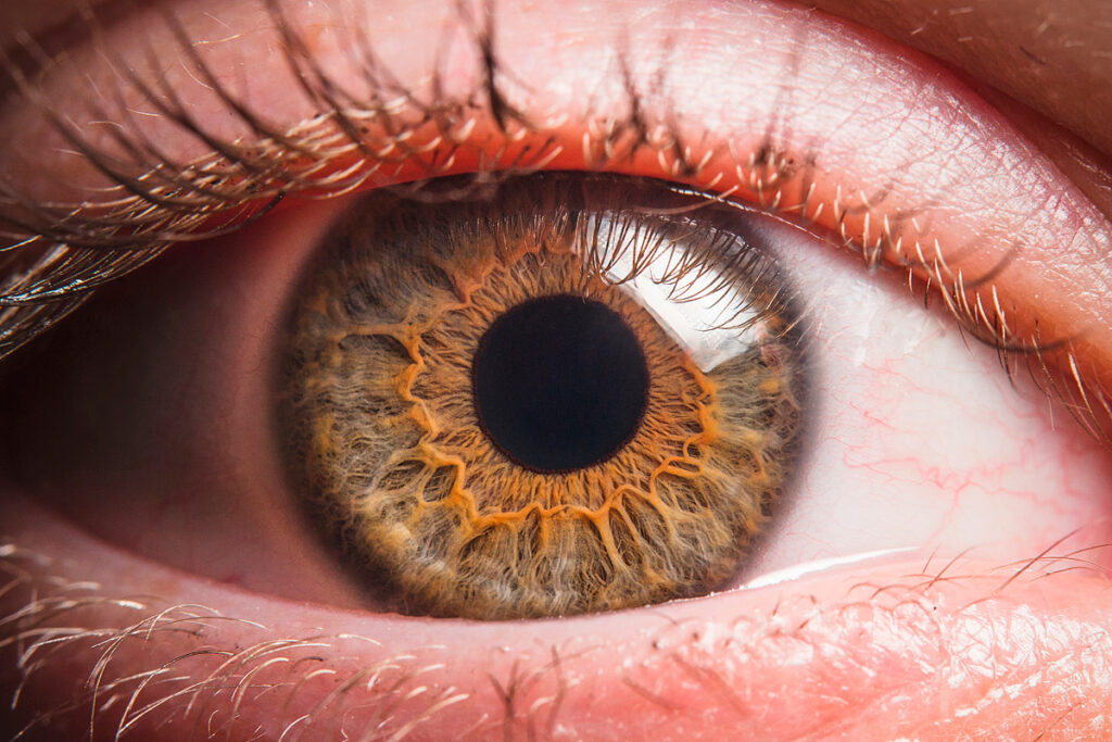
The SARS-CoV-2 coronavirus caused the COVID-19 pandemic. It has changed the lives of billions of people, infected more than 150 million, killed more than 3 million people. Understanding the mechanisms of infection and all the ways the virus enters the body will help minimize the spread of coronavirus infection.
In May 2021, an international group of scientists from the United States and Denmark proved the possibility of direct infection with coronavirus through the eyes. Experts have shown that the cells of the eye surface and the back of the eye have the necessary mechanism for developing infection and are easily infected under experimental conditions. In a series of laboratory experiments, experts have found that the most susceptible to infection is the limbus cells – the transition zone between the cornea of the eye and the sclera.
The scientists also demonstrated the vital role of the TMPRSS2 serine protease, which, together with the ACE2 cell receptor, ensures the virus’s penetration into the cell and its cleavage into individual proteins. Suppression of TMPRSS2 serine protease completely eliminates infection before cell infection with coronavirus.
Medical significance of the study
The medical community has not yet reached a consensus on protecting the eyes from coronavirus infection. Several scientists believe that the risk of infection through the eyes is exaggerated. On the other hand, in 1-5% of patients with COVID-19, the coronavirus is detected in the tear fluid. Also, a study of more than 25,000 patients revealed a relationship between eye protection and a decrease in the level of infection.
Scientists explain that even 1% of cases of eye complications in COVID-19 should not be neglected – after all, this is more than 1 million people around the world.
Research design
The study used donated eye tissue from three patients who died from a coronavirus infection. To determine how healthy cells react to infection, the scientists also used the eye tissues of 4 people who died from other diseases and were not infected with SARS-CoV-2. The donor organs were obtained from the eye bank, New York (USA), within 24 hours after the death of the patients. The experiments studied the tissues of the anterior and posterior segments of the eyes: sclera, cornea, limbus, retina, choroid, iris.
To obtain more complete data, the scientists used as a model a cell culture of eye organoids obtained from human embryonic stem cells. This model eliminated the dependence of the study on the donated eyes of deceased patients and facilitated the study of SARS-CoV-2 in eye tissues.
Study results
Assessment of infection of the eye tissue of donors who died from coronavirus infection
First, scientists examined the anterior segments of the eyes of people who died from COVID-19: the sclera, limbus, cornea. The SARS-CoV-2 spike protein was found in the ocular epithelium of all three donors. However, it was not in the corneal stroma.
The SARS-CoV-2 nucleocapsid protein was also present in the cells of the eye surface. Moreover, most of it was in the limbal region.
Comparison of eye tissue types by susceptibility to SARS-CoV-2
Then the specialists assessed which areas of the eye are most susceptible to infection. To do this, scientists infected healthy cells of six eye tissues with the SARS-CoV-2 coronavirus: the cornea, limbus, sclera, iris, retina, and vascular membrane.
The coronavirus successfully multiplied in the cells of the cornea, sclera, limbus and retina. The expression of SARS-CoV-2 was not detected in the vasculature and iris. The quantitative assessment showed that limbal cells are significantly more infected than corneal or sclera cells.
The ACE2 receptor analysis, through which the virus enters the cell, and the TMPRSS2 serine protease, which cleaves the virus, showed that they are most strongly expressed in the limbal region.
Evaluation of the role of serine protease TMPRSS2 and ACE2 receptor in the development of coronavirus infection
To test the dependence of the infection on serine proteases, the scientists suppressed their activity in limbus cells using NP-tosyl-L-phenylalanine chloromethyl ketone. As a result, the pretreatment of the cells with this compound completely eliminated the infection. Experts believe that, perhaps, the limbus is most susceptible to infection since it contains corneal and conjunctival stem cells.
To understand why the limbus is most susceptible to infection, scientists studied the genes of limbal cells compared to corneal cells. It was found that the genes ACE2 and serine proteases TMPRSS2, TMPRSS4, TMPRSS11E, which are associated with SARS-CoV-2 infection, are most active in the limbus. Scientists also found that the morphogenesis of keratinization of the epithelium is characteristic of limbus cells. Similar morphogenesis of keratinization is characteristic of primary epithelial cells of the lungs when infected with SARS-CoV-2.
Further genetic analysis showed that many of the same genes are activated in infected limbal cells and infected cells of the bronchial epithelium. Moreover, the genes responsible for normal limbal functions are suppressed.
Cultivation of multiple lines of eye organoids
The scientists continued their research on an organoid model of the eye obtained from human pluripotent stem cells. This model eliminated the dependence of the study on the donated eyes of deceased patients and facilitated the study of SARS-CoV-2 in eye tissues.
After 7 weeks of stem cell differentiation, the researchers obtained cells of the developing ocular field, the ectoderm of the ocular surface, retinal epithelium, dorsal ocular cup and periocular mesenchyma. Genetic analysis showed that the cultures developed towards corneal cells, epidermal cells, conjunctiva and limbus. However, these populations were immature.
Analysis of the model for the presence of coronavirus input factors
Then, in the obtained organoids, the scientists assessed the presence of infection factors with the SARS-CoV-2 coronavirus. It turned out that the ACE2 receptor is expressed in 74% of the cells of the ocular surface, especially in the population of conjunctival and limbus cells. Moreover, the same genes were activated in the immune responses of the model cells as in mature cells of the multilayer epithelium expressing ACE2.
Then the scientists studied the expression of the serine protease TMPRSS2 in the organoids of the eye. TMPRSS2 expression was detected in 82% of ectoderm cells on the surface of the eye. Moreover, the organoid of the conjunctiva and limbus contained the most significant number of such cells. As a result, the scientists suggested that the organoid model is suitable for studying eye infection, and the cells may be susceptible to SARS-CoV-2.
Assessment of the infectivity of conjunctival and limbal cells
To quantify virus replication dynamics, specialists infected the organoids of the conjunctiva and limbus with coronavirus and incubated them for 24 hours. The number of infectious SARS-CoV-2 virions has increased by 100 times. The viral load reached a plateau after 24 hours and persisted for three days.
Since the ACE2 receptor and the TMPRSS2 serine protease were more common in the organoids of the conjunctiva and limbus, the scientists assumed that these cells would be infected in the first place. The analysis of viral RNA showed that the spike protein SARS-CoV-2 is most often expressed in the organoids of the cornea and limbus. Moreover, the limbus was the most infected. That confirms the previous observation that the limbus cells of postmortem donors are more susceptible to infection than the cells of other eye tissues.
Evaluation of the antiviral response of infected cells of the eye model
Then the scientists evaluated the antiviral response of the infected organoids of the cornea and limbus. Interferons of types I and III (IFN-I, IFN-III) serve as the first line of defense against viral infections, but successful viruses can counteract the interferon response. The SARS-CoV-2 genome encodes several proteins that suppress the cellular antiviral response, including the interferon response.
The researchers compared the expression of interferon-stimulated genes (ISG) of healthy and infected cells. ISG activity was lower in infected limbic organoids than in infected corneal organoids but higher than background expression in healthy cells. These data confirm the ability of SARS-CoV-2 to counteract the work of interferon-stimulated genes in cells.
Thus, scientists have shown that the SARS-CoV-2 coronavirus captures the protein synthesis system in an infected cell to replicate its genome at the expense of normal cell functions.
Conclusions
SARS-CoV-2 eye infection occurs mainly in the limbus, which serves as a reservoir of new viral virions.
The eye surface cells are very susceptible to SARS-CoV-2 infection, and therefore, eye protection is required as a potential site of infection.
The serine protease TMPRSS2 is critical for infecting a cell with a coronavirus. Suppression of TMPRSS2 completely eliminates the infection.
Source
S ARS-CoV-2 infects human adult donor eyes and hESC-derived ocular epithelium