Cardiovascular complications of COVID-19
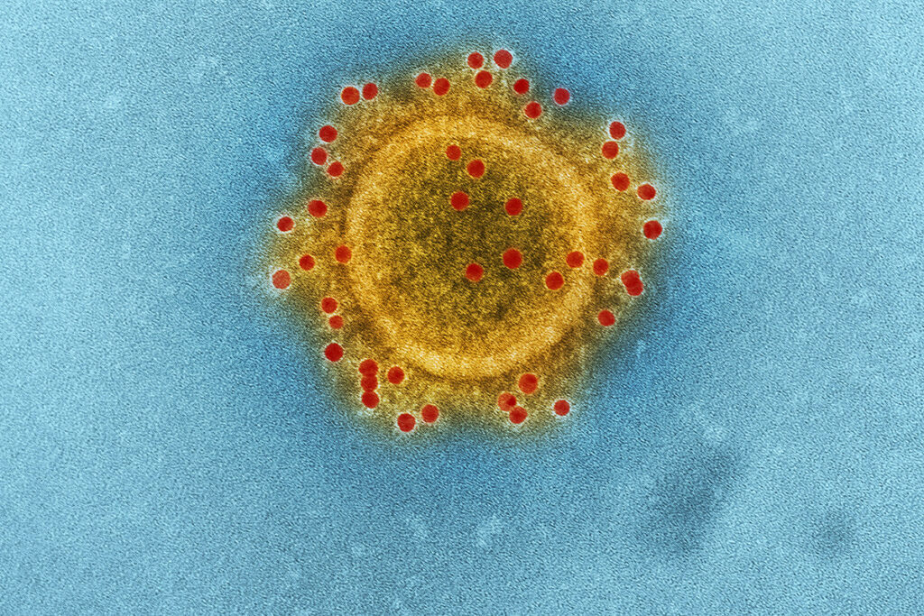
Usually, coronaviruses cause symptoms from the respiratory system. However often, viruses damage other organs as well. The main target of the SARS-CoV-2 coronavirus is the lungs. However, cardiovascular complications, including the progression of pre-existing and the emergence of new diseases, increased the number of deaths from the coronavirus disease COVID-19.
The most common cardiovascular complications after SARS-CoV-2 infection are myocardial damage, arrhythmias, and heart failure.
Troponin I is a protein that regulates heart contractions. It is released into the blood during myocardial infarction. Myocardial injury characterized by elevated serum cardiac troponin I levels or electrocardiogram abnormalities has been associated with increased mortality in COVID-19 patients.
The SARS-CoV-2 coronavirus can cause acute coronary syndrome even in the absence of systemic inflammation. Hospitalized patients with COVID-19 develop cardiac arrhythmias, including ventricular tachycardia and atrial fibrillation. Progressive left ventricular dysfunction, and general symptoms resembling heart failure were also observed in a significant number of patients. At the beginning of the outbreak, these symptoms were mainly observed in critically ill patients with COVID-19. However, recent studies report that cardiac symptoms are also observed in mild and even asymptomatic cases of COVID-19.
The mechanisms of cardiovascular complications of COVID-19 are still unclear. In a lung infection, an uncontrolled release of inflammatory cytokines, called a cytokine storm, can cause damage to multiple organs, leading to organ failure and the progression of pre-existing cardiovascular diseases. Moreover, COVID-19 alters blood clotting, which can also cause coronary heart disease. Finally, SARS-CoV-2 can directly infect heart cells by penetrating them through the ACE2 protein. This protein is expressed in various human body tissues, including heart cells, cardiomyocytes, and pericytes.
Several studies have identified the SARS-CoV-2 genome in the heart and reported signs of viral myocarditis in patients with COVID-19, including asymptomatic cases. In vivo and in vitro studies using adult human cardiomyocytes and cardiomyocytes derived from human pluripotent stem cells (hpSC-CM) have shown that SARS-CoV-2 can infect cardiomyocytes.
Scientists at the University of Washington in Seattle, USA, investigated whether human cardiomyocytes with the SARS-CoV-2 coronavirus lead to impaired cardiac function and whether other types of heart cells are susceptible to SARS-CoV-2. The researchers used cardiomyocytes derived from human pluripotent stem cells (hpSC-CM) and smooth muscle cells (hpSC-SMC) for the study.
Proteins that promote the penetration of coronavirus have been found in the muscle cells of the heart
Susceptibility to SARS-CoV-2 infection depends on the expression of both the ACE2 receptor and various proteases – enzymes that break down proteins. Proteases can cleave the coronavirus’s spike docking protein, which leads to the release of the SARS-CoV-2 genome into the cell’s internal environment.
The ACE2 protein was found in 9% of cardiomyocytes derived from embryonic stem cells (hESC-CM). That indicates a low or temporary expression of ACE2. Nevertheless, most of the cells expressed moderate to high protease levels: CTSB-71%, CTSL-46%. These proteases are often expressed in conjunction with ACE2. Although the TMPRSS2 protease can also mediate virus entry, this protease has not been detected in either hESC-CM or adult heart cells. On the other hand, the PIKFYVE and FURIN proteases were widely expressed in hESC-CM. It indicates that the mechanism of SARS-CoV-2 penetration into cardiomyocytes may differ from the TMPRSS2-dependent mechanism of penetration into lung epithelial cells.
The ACE2 protein was detected in hpSC-CM. The levels of ACE2 were comparable to the levels in the epithelial cells of the kidneys of primates, for which the possibility of infection with the SARS-COV-2 coronavirus has been proven.
These results show that human stem cell-derived cardiomyocytes (hpSC-CM) express proteins that may make them susceptible to SARS-CoV-2 infection.
SARS-CoV-2 can infect the heart’s muscle cells and multiply in them using the ACE2 receptor
The highest levels of ACE2 were found in hpSC-CM cardiomyocytes. Scientists have tested whether SARS-CoV-2 can infect hpSC-CM and multiply in them.
Infection of cardiomyocytes
The scientists incubated high-purity hpSC-CM with the SARS-CoV-2 coronavirus and tested the results for different amounts of viral particles per cell (MOI):
- MOI – virus spreading inside the cells and secondary infection of others.
- 5 MOI – infection of all susceptible cells.
In both cases, the coronavirus affected hpSC-CM cardiomyocytes. As expected, at 5 MOI, the effect was faster. The highest MOI led to the cessation of heartbeats and the appearance of signs of the death of heart muscle cells within 48 hours after infection. The effects were more pronounced for the female cardiomyocyte cell line.
Replication of SARS-CoV-2 in cardiomyocytes
To find out whether SARS-CoV-2 can replicate in cardiomyocytes, the scientists quantified extracellular viral particles and intracellular viral RNA:
At 5 MOI, virus replication was stable from 24 to 72 hours after infection, followed by a sharp decrease as cells died.
At 0.1 MOI, virus replication occurred with a noticeable increase in viral particles and RNA from 48 to 72 hours after infection.
Female cardiomyocytes were more susceptible to SARS-CoV-2 replication than male cardiomyocytes.
Unlike cardiomyocytes derived from human pluripotent stem cells (hpSC-CM), SARS-CoV-2 did not affect cardiomyocytes derived from smooth muscle cells (hESC-SMC) even at the highest MOI 72 hours after infection. The number of extracellular viral particles and intracellular viral RNA in hpSC-SMC was more than two orders of magnitude lower than that observed for hpSC-CM. It highlights that the SARS-CoV-2 coronavirus infects and replicates precisely in cardiomyocytes derived from human pluripotent stem cells.
The role of ACE2 during SARS-CoV-2 cardiomyocyte infection
Scientists have tested what will happen under the influence of SARS-CoV-2 with cardiomyocytes without the ACE2 protein. Even at 5 MOI, the absence of ACE2 prevented cell death. The coronavirus was detected 48 hours after infection at 0.1 MOI only in wild-type cardiomyocytes but not in cardiomyocytes without ACE2. These results show that the infection of SARS-CoV-2 cardiomyocytes is mainly dependent on the expression of ACE2.
SARS-CoV-2 virus entry, replication, and exit
Electron microscopic studies have demonstrated the virus’s penetration through the direct fusion of the viral envelope with the cell membrane and through endocytosis – the capture of the viral particle by the cell, during which the cell envelops the viral particle with the membrane and swallows it.
After the virus entered the cell, its replication was observed in two-membrane structures inside the cell. For the mass release of mature viral particles, the virus captured the lysosome – the part of the cell that can secrete its contents into the extracellular environment.
SARS-CoV-2 reprograms cellular processes
Scientists have investigated what changes the coronavirus causes in cells. SARS-CoV-2 can reprogram cellular gene expression to promote its replication.
The coronavirus suppresses the genes responsible for mitochondrial function and energy production. Mitochondria are the parts of the cell that are responsible for converting nutrients into energy – ATP. ATP is necessary to transmit nerve impulses, muscle contraction, heat formation, and metabolic processes maintenance. SARS-CoV-2 can inhibit the accumulation of ATP in mitochondria and shift the metabolism from oxidative to glycolytic, contributing to replicating the virus.
The innate immune system recognizes pathogenic microorganisms that have entered the body with pattern recognition receptors (PRRS). PRRS identifies a set of nucleic acids and virus replication products that are not present in the body’s original cells and triggers an early immune response. Activation of innate immunity leads to the production of type I and type III interferons, and interferon triggers antiviral protection.
Scientists analyzed the interferon response after infection with the coronavirus hpSC-CM cells. 48 and 72 hours after infection, the expression of genes encoding the signal protein interferon increased in the cells. A more substantial effect was observed in the female cardiomyocyte cell line. These results indicate that SARS-CoV-2 activates innate immunity and interferon response in hpSC-CM cardiomyocytes.
SARS-CoV-2 creates a risk of arrhythmias in heart muscle cells
The researchers investigated whether SARS-CoV-2 infection interferes with hpSC-CM function. Unlike the previous experiment on infecting sparse cardiomyocyte cultures with coronavirus, in the current investigation, hpSC-CM cells were seeded at a high density to evaluate the electrophysiological properties reliably. In this experiment, there were no tremendous cytopathic effects. However, viral RNA and viral particles were still detected, showing that infection also occurred in high-density cultures.
Infection with SARS-CoV-2 quickly led to decreased ripple frequency, decreased amplitude of the depolarization burst, and decreased electrical conductivity rate. In the female cardiomyocyte line, an increase in the interval between depolarization and membrane repolarization (FPD) was observed both in cultures with spontaneous beating and in cultures with electrical stimulation. The gap between depolarization and repolarization is an in vitro surrogate of the QT interval measured by an electrocardiogram.
Overall, disturbances in the generation and propagation of electrical signals were significant even in the absence of extensive cell death. It suggests that SARS-CoV-2 infection in cardiomyocytes may provoke arrhythmias observed in 20% of patients with COVID-19.
SARS-CoV-2 infection reduces the strength of contractions in artificial heart tissues
The scientists evaluated the contractile properties of hpSC-CM using 3D-EHT three-dimensional engineered heart tissues, tracking their contractile behavior by measuring the magnetic field. Cardiomyocytes in 3D-EHT were infected at 10 MOI SARS-CoV-2, and their reduction was analyzed within a week.
Viral replication was comparable to reproduction in two-dimensional structures from previous experiments, highlighting the possibility of infecting heart muscle cells with coronavirus.
The maximum strength of contractions in infected tissues decreased within 72 hours after infection. Within 144 hours of infection, the contractions continued to decline to less than 25% of the original strength. The density of cardiomyocytes progressively decreased, while the cells became more rounded and less aligned along the longitudinal axis of the 3D-EHT. Together, this can help to reduce the strength of the heart rate.
In infected 3D-EHT cells, the expression of the sarcomeric genes MYL2 and MYH6 was reduced. A sarcomere is a contractile unit of a muscle. Reduced expression of sarcomeric genes may correlate with changes in the structure of the sarcomere.
A significant violation of the contractile properties of 3D-EHT demonstrates that SARS-CoV-2 infection affects the mechanical function of cardiomyocytes in vitro. Similar mechanisms may contribute to cardiac dysfunction in patients with COVID-19.
Conclusions
SARS-CoV-2 can directly infect cardiomyocytes. Regardless of inflammation or changes in blood clotting, it can disrupt the electrophysiological and contractile properties of cardiomyocytes and cause their death. Cardiomyocytes, but not smooth muscle cells, express ACE2, making them susceptible to SARS-CoV-2 infection. ACE2 is a crucial factor for SARS-CoV-2 entry into cardiomyocytes.
The interferon response, which is part of the innate immune response activation, usually occurs within a few hours after a viral infection. After entering the cell, the coronavirus is located in two-membrane vesicles to replicate and hide from the immune system effectively. Therefore, the activation of the genes responsible for interferon production was observed only in the late stages of infection with SARS-CoV-2. The fact that cardiomyocytes infected with SARS-CoV-2 exhibit a delayed interferon response may promote virus replication to high levels.
To prevent systemic inflammation, patients with COVID-19 are usually treated with steroids. However, treatment aimed at preventing infection, viral replication, and direct damage to heart cells by the SARS-CoV-2 coronavirus and restoring heart function can prevent long-term cardiovascular complications.


