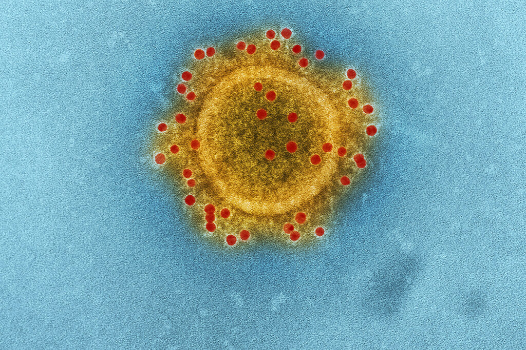
The SARS-CoV-2 coronavirus can cause a wide range of diseases, from mild upper respiratory tract disease to severe respiratory failure, which requires mechanical ventilation. At risk for severe COVID-19 are people with chronic lung diseases such as severe asthma and chronic obstructive pulmonary disease (COPD).
However, COPD is less common among hospitalized people with COVID-19 than diseases such as hypertension and diabetes. Therefore, it can be assumed that people with COPD are better protected from contracting coronavirus.
But, if a person with COPD is still infected with the SARS-CoV-2 coronavirus, he has a higher risk of severe COVID-19 and death. The risk is also increased in people with severe asthma and smokers.
Inhaled corticosteroids (ICS) are drugs used to treat respiratory conditions such as COPD and asthma. These drugs protect against flare-ups, reduce the risk of contracting the virus, suppress the inflammation caused by the virus, and prevent symptomatic manifestations.
Nevertheless, studies have shown that ICS suppresses the innate immune response to rhinovirus and influenza infection, which leads to increased viral replication, although other studies have recorded a protective effect of ICS against seasonal coronavirus 229E and SARS-CoV-2.
These facts raise two questions:
- Do ICS generally have a protective or detrimental effect on the immune response to SARS-CoV-2?
- Does ICS protect or expose patients with COPD against COVID-19?
How COPD, Asthma, and Smoking Affect the Expression of the ACE2 Coronavirus Receptor
The two main proteins by which SARS-CoV-2 penetrates the mucous membrane of the respiratory tract and causes infection are ACE2 and TMPRSS2. The SARS-CoV-2 coronavirus uses the cellular receptor ACE2 to attach to a cell. However, for the viral particle to enter the cell, it must be processed by the TMPRSS2 protein.
In the epithelium of the respiratory tract of smokers and patients with COPD, the expression of ACE2 is increased. It is believed to predispose them to a poor outcome from COVID-19. In asthma, on the other hand, ACE2 is suppressed. This effect can be associated both with the action of type II cytokines and ICS use.
Inhaled Corticosteroids Suppress the Expression of Type I Interferon and ISG
Antiviral mechanisms regulate ACE2 expression in the airways. In the nasal epithelium cells, ACE2 is expressed in conjunction with immune genes involved in interferon signaling. In addition, scientists recently found out that ACE2 is an interferon-stimulated gene (ISG) in the cells of the human respiratory epithelium.
ICS suppresses epithelial expression of type I interferons and ISG in several in vitro and in vivo models of COPD. The question arose before scientists: could ICS-induced suppression of interferon lead to a decrease in the level of ACE2 in the lungs and, thus, affect the susceptibility to SARS-CoV-2 in patients with chronic lung disease?
Study of The Relationship Between Inhaled Corticosteroid Use and ACE2 Expression
British scientists conducted a study on 40 patients with COPD in a clinically stable condition. The patients were divided into two groups:
- those who have used ICS either in a single-agent inhaler or in combination with bronchodilators;
- those who did not use ICS.
Scientists obtained primary bronchial epithelial cells (BEC) from 6 patients with COPD and 6 healthy non-smokers from the control group. Cells were treated with 10 nMol / L corticosteroid fluticasone propionate (FP) or dimethyl sulfoxide, a carrier substance (VEH in figures).
For the study, the researchers also used Ifnar- / – mice with impaired interferon signaling. Mice were injected intranasally with corticosteroids FP, budesonide, or beclomethasone in the form of a solution containing 20, 6.7, or 2 μg of the active ingredient, respectively.
Mice were selected for analysis 8, 24, 48, or 96 hours after FP administration. In some experiments, 2 hours after FP administration, mice were injected intranasally with 50 μl of saline containing 104 recombinant interferon-beta (IFN-beta) units.
Pulmonary ACE2 Expression is Decreased in COPD Patients Taking Inhaled Corticosteroids
Studies indicate that the expression of ACE2 mRNA in sputum is reduced in patients with bronchial asthma taking ICS.
Scientists selected 18 patients, each from those who used ICS and those who did not use – a total of 36 patients. Expression of ACE2 mRNA in sputum cells was detected in 22 of 36 patients with COPD (61.1%). As in asthma, ACE2 expression was significantly reduced in patients taking ICS than those who did not (Fig. 1, A).
Expression by sputum cells of the TMPRSS2 protein, which SARS-CoV-2 uses to penetrate the mucous membranes, was found in all subjects, with no significant difference between those who took ICS and those who did not (Fig. 1, B).
Fig. 1. Expression of the ACE2 and TMPRSS2 genes in the sputum of patients with COPD

Likewise, the alternative SARS-CoV receptor CD147 (basigin gene, BSG) was found in all subjects, with no difference observed between those who took ICS and those who did not (Figure E2).
Fig. E2. BSG gene expression in sputum of patients with COPD

Inhaled corticosteroid administration suppresses pulmonary ACE2 expression in mice
To separate cause from effect, the researchers tested in mice whether experimental pulmonary administration of ICF to FP mice suppressed ACE2 expression.
A single injection of 20 µg FP to mice (Fig. 2, A) significantly suppressed the expression of ACE2 mRNA in the lungs after 8 hours. 8 o’clock is the point in time at which significant activation of the glucocorticoid receptor (GR) occurs.
This effect persisted 24 hours after administration but disappeared after 48 hours (Fig. 2, B).
Suppression of ACE2 by FP occurred in a dose-dependent manner with a loss of suppression at a 10-fold lower concentration (2 μg) (Fig. 2, C) – a dose at which the effect of GR activation is also lost.
Consistent with the effects observed in human sputum, FP administration did not affect the expression of TMPRSS2 or BSG in mouse lungs.
Scientists observed a similar suppression of ACE2 mRNA expression in the lungs when administered with 20 μg of other widely used ICS – budesonide and beclomethasone. It means that the effect of ICS on ACE2 does not depend on the drug class (Fig. 2, D).
Fig. 2. The introduction of ICS suppresses the expression of ACE2 in the lungs of the mouse

The ACE2 Suppression by FP is Associated with Suppression of Type I Interferon
ACE2 is an interferon-stimulated gene in the airways and is co-expressed with type I interferon (IFN-I) genes. It means that IFN-I may be the primary regulator of pulmonary ACE2 expression.
Since the expression of IFN-beta in the airways is reduced in patients with COPD taking ICS, the researchers hypothesized that the decrease in the expression of ACE2 by FP might be due to its suppressive effect on type I interferon signaling. Accordingly, administration of recombinant IFN-beta (Fig. 3, B) can reverse FP-induced suppression of ACE2 (Fig. 3, C). This fact indicates that the effect of FP on ACE2 expression is functionally related to the fact that FP suppresses IFN-I.
Fig. 3. Suppression of ACE2 by ICS is associated with suppression of type I interferon

Basal ACE2 expression in Ifnar- / – mice
To confirm the functional significance of type I interferon in the regulation of pulmonary ACE2, the researchers assessed the basal levels of pulmonary expression in mice with deficient interferon signaling (Ifnar- / -).
Compared to wild-type control mice, Ifnar- / – mice had a small but statistically significant decrease in ACE2 mRNA expression in the lungs (Fig. 4, A), with a concomitant trend towards reducing the level of ACE2 protein in the lungs (Fig. 4, B). These observations support the critical regulatory role played by IFN-I signaling in the pulmonary expression of ACE2.
Fig. 4. Mice deficient in type I interferon receptors have reduced expression of ACE2 in the lungs

Inhaled corticosteroids suppress ACE2 expression in bronchial epithelium
The ACE2 receptor is expressed mainly in the nasal and bronchial epithelium and is absent from immune cells. FP suppresses the immune system of the pulmonary epithelium.
Scientists evaluated whether ACE2 expression was inhibited after ICS administration in cultured primary bronchial epithelial cells (BEC) obtained from COPD patients (Fig. 5, A).
Experiment Results:
- Basal ACE2 expression was increased approximately 3-fold in BECs from COPD patients compared to cells from healthy non-smokers (Fig. 5, B).
- Administration of FP at a clinically significant concentration of 10 nMol / L inhibited ACE2 expression by 75% (Fig. 5, C).
- FP administration to COPD BEC did not affect TMPRSS2 expression (Fig. E4).
Fig. 5. ACE2 expression is increased in COPD and is suppressed by the administration of fluticasone in cultured bronchial epithelial cells (BEC)

FP suppresses ACE2 expression in the lungs of COPD mice
The researchers used a mouse model of emphysema caused by elastase, an enzyme that breaks down protein. This model reproduces many of the characteristics of the disease in humans.
The expression of ACE2 in the lungs was measured 10 days after the administration of elastase. It was the moment when the signs of COPD-like disease were established and after another seven days.
In mice treated with elastase, the expression of ACE2 in the lungs increased 5-fold after 10 days, with a further increase of more than 15-fold after 17 days (Fig. 6, B).
The administration of a single dose of FP 10 days later attenuated the expression of ACE2 mRNA in the lungs (Fig. 6, C) and decreased the concentrations of the ACE2 protein measured after 24 hours (Fig. 6, D), but did not affect TMPRSS2.
These results confirm that ICS suppresses ACE2 in an in vivo model of COPD-like disease.
Fig. 6. Expression of ACE2 is increased in the mouse model of COPD and is suppressed by the administration of fluticasone

Conclusions
The ACE2 receptor, which facilitates the entry of SARS-CoV-2 into the airways, is activated in COPD. The introduction of ICS decreases the pulmonary expression of ACE2. The decrease in ACE2 expression is since ICS suppresses type I interferon. These data indicate that the use of ICS therapy alters the expression of the SARS-CoV-2 entry receptor, thus altering COVID-19 susceptibility in COPD patients.
The exact role of ACE2 in the pathogenesis of COVID-19 has not been fully characterized. However, some facts indicate the critical role of ACE2 in the penetration of the virus and the pathogenesis of SARS-CoV:
- In SARS-CoV-1, higher expression of ACE2 increases susceptibility to infection in vitro.
- In animal studies, ACE2 overexpression enhances SARS-CoV-1 penetration, anti-ACE2 antibodies can block viral invasion, and ACE2-deficient mice have a decrease in pulmonary pathology.
- ACE2 is expressed primarily in the goblet cells of the nose and type II pneumocytes in the airways. It is activated in people at risk of serious illness: the elderly and patients with diabetes.
Suppression of ACE2 can worsen disease outcome:
- In murine models of aspiration pneumonia and sepsis, genetic deletion of ACE2 exacerbates acute lung injury. The introduction of recombinant ACE2 partially eliminates this effect.
- ACE2 breaks down angiotensin II, which can drive the production of pro-inflammatory cytokines. It can be harmful in the context of hyperinflammation associated with severe COVID-19.
COPD may also be associated with possible protection against coronavirus infection and, if infected, with an increased propensity for severe COVID-19. ICS suppresses type I interferon, leading to increased viral replication and the likelihood of secondary bacterial infection. The introduction of recombinant IFN-beta in combination with FP can reverse the suppression of ACE2 expression.
Understanding the mechanism by which ICS suppresses ACE2 expression is essential in determining how this effect can be used as a protective factor or reversed if deleterious.