Content
- Estimating a safe dose of alcohol for the brain depends on the drink type.
- Moderate alcohol consumption affects brain function and cognitive abilities.
- Study of the mechanisms of the negative effect of alcohol on the brain.
- How Alcohol Promotes Iron Accumulation, Disrupts Iron Balance and Stimulates Cell Death.
There is a myth that moderate alcohol consumption does not affect the body. It even increases the level of creativity and improves the quality of thinking.
However, scientists from the universities of Oxford and Cambridge (UK) refute this strong opinion. Scholars’ research studies and evidence:
- Measurement of alcohol doses does not exist;
- there are no safe or healthy alcoholic beverages for the brain: wine, beer, spirits, and their combinations all cause degenerative processes in the brain, resulting in dementia used to Alzheimer’s and Parkinson’s;
- alcohol poisoning of almost all areas of the brain, the gray and white matter of the brain accumulates, and the activity of neurons is delayed;
- Even with moderate alcohol consumption, cognitive functions decrease, and memory deteriorates.
The negative effect of alcohol on the brain is caused by a violation of iron metabolism in the body. Carbohydrate alcohol synthesizes hepcidin, a hormone, due to the intake of which the body absorbs iron through the intestines. Then excess iron is deposited in various organs, including the brain and nerve cells. As a result, the volume of the brain increased. This process is called ferroptosis – cell death due to oxidative stress caused by iron.
Scientists note that the negative effect of alcohol on the brain is much higher than before. They are calling for rethinking recommendations for alcohol consumption in light of its observation of brain health.
Research on Calculating Blood Alcohol Concentration Measurement Depending on The Type 0f Spirit
Study Design
In this experiment, scientists sought to determine the minimum amount of alcohol that does not harm the brain.
First, for 5 years (2006-2010), scientists interviewed participants about the amount of alcohol consumed, the regularity, and the type of spirit drink. Then there was a pause in the study. And then, for another 7 years (2014-2020), all participants had magnetic resonance imaging (MRI) of various brain areas and conducted cognitive tests.
Using MRI, scientists determined the brain’s health and structural and functional changes. And with the help of cognitive tests, it became clear how these changes are reflected in a person’s daily life.
The study involved 25,378 people, the average age was 55 years.
Research results
The dose of alcohol consumed directly proportionally reduces the volume of gray matter in the brain and lowers its density. Gray matter is all nerve cells, auxiliary cells (glia), and brain capillaries. The gray matter of the brain is responsible for mental and muscular activity. For example, it provides thinking, emotions, speech, memory, sight, hearing, taste, and touch.
According to the results of the MRI, it was found that alcohol has the most substantial effect on the thalamus, which is the part of the brain responsible for attention concentration, regulates the level of consciousness, controls sleep and wakefulness, and transmits signals from the senses.
An MRI of the brain shows the average degree of influence of alcohol on the density of gray matter. Scientists emphasize that this is only the influence of alcohol, and other factors were not taken into account when creating the image: head size, age, gender, cholesterol (HDL, LDL), blood pressure, diabetes, smoking, physical activity, body mass index (BMI), the presence of depression.

Red-yellow areas show a decrease in the density of gray matter.
Image source: https://www.medrxiv.org/content/10.1101/2021.05.10.21256931v1.full
Alcohol dose-dependently worsens the integrity of the brain’s white matter, reduces its density, disrupts the functional connectivity of different parts of the brain, leads to a shortage of water in neurons, and increases the amount of water in the intercellular space in the opposite, which disrupts the microstructure. Violation of the functional connectivity of brain regions reduces creativity and the ability to solve complex problems.
White matter is the long process of nerve cells (axons) covered with myelin (white fat). The brain’s white matter is responsible for the speed of signal transmission between nerve cells. Also, white matter connects the right and left hemispheres of the brain. This connection between the hemispheres, called the corpus callosum, turned out to be the most vulnerable part of the white matter to the adverse effects of alcohol. Deterioration of the microstructure of the corpus callosum is an indicator of vascular dementia, as a result of which the speed of movement and the rate of information processing decrease, as well as the executive functions of the brain deteriorate.
An MRI of the brain shows areas of violation of the integrity of the white matter caused by alcohol. As in the previous picture, this is solely the influence of alcohol, without taking into account other individual factors.

The red areas show the brain’s most severe white matter destruction.
Image source: https://www.medrxiv.org/content/10.1101/2021.05.10.21256931v1.full
Of all the negative factors affecting the brain, alcohol is the most significant. High blood pressure, obesity, high cholesterol, and smoking negatively impact brain health. But the adverse effect of alcohol, for example, is 4 times higher than smoking. Moreover, other negative factors increase the negative impact of alcohol on the brain.
Non-drinkers were 5.2% of all study participants. Those who have never drunk – are 691 people. Those who have stopped drinking alcohol – 617. Those who currently continue to drink alcoholic beverages – 24,069 people. Scientists conclude that alcohol consumption is prevalent.
The negative effect on the brain does not depend on the type of alcoholic beverage. Wine, beer, and spirits – all equally harmful to the brain, and that confirm the hypothesis that ethanol, and not other compounds in alcoholic drinks, leads to neuron damage.
There is no minimum dose of alcohol that is safe for brain health.
Effect of Moderate Alcohol Consumption on Brain Function and Cognitive Abilities
Research design
In a series of experiments conducted over 30 years (1985-2015), scientists were looking for answers to the question — how does moderate alcohol consumption affect the structure and function of the brain: positively, negatively, or not?
At intervals of 5 years, scientists interviewed people about the weekly level of alcohol consumed, noted changes in social status, did medical tests, and conducted cognitive tests. At the end of the study, all participants had an MRI of the head and studied the density of gray matter, the microstructure of the brain’s white matter, and changes in the hippocampus. The hippocampus is an area of the brain responsible for long-term and spatial memory, forms emotions, provides concentration and attention retention, and implements foresight and intuition mechanisms.
527 people participated in the study, and the average age was 43.
Research results
The degree of degradation of the hippocampus is directly proportional to the dose of alcohol consumed. The myth about the protective effect of small amounts of alcohol (1-8 grams of ethanol per week) on the brain has not been confirmed. The biggest atrophy of the hippocampus was in people who consumed more than 240 grams of ethanol per week — 5.2 times higher than in non-drinkers. The degree of atrophy in moderate drinkers (56-104 grams of ethanol per week) was 3.2 times higher than in non-drinkers. Scientists remind us that damage to the hippocampus is an early and specific indicator of Alzheimer’s disease.
The researchers recruited three clinicians to test a previously uncharacteristic dose-dependent relationship between alcohol and hippocampal degradation. Each doctor, independently of the others, conducted an independent assessment of the degree of atrophy. The conclusions of the doctors coincided with the results that the scientists received. As a result, the researchers could deduce the relationship between the weekly level of alcohol consumed and the degree of degradation of the hippocampus. Detailed results of the experiment are given in the table:
| Ethanol consumption per week | Atrophy rate of the RIGHT part of the hippocampus | Atrophy rate of the LEFT part of the hippocampus |
| Men | ||
| 0 — <1 | — | — |
| 1 — <7 | 1,6 | 1,7 |
| 7 — <14 | 1,7 | 1,5 |
| 14 — <21 | 3,2 | 2,0 |
| 21 — <30 | 3,9 | 2,2 |
| 30 and more | 5,2 | 6,3 |
| Women | ||
| 0 — <1 | — | — |
| 1 — <7 | 1,1 | 0,8 |
| 7 — <14 | 3,1 | 2,0 |
| 14 — <21 | 4,2 | 6,2 |
| 21 — <30 | 1,1 | 0,4 |
| 30 and more | — | — |
Alcohol reduces the density of the gray matter of the brain. Scientists have found the most remarkable changes in the hippocampus and the amygdala. The amygdala of the brain processes signals from the senses, forms emotions and emotional memory and participates in decision-making. Dysfunction of the amygdala leads to bipolar disorder and dulls fear.
Alcohol violates the integrity of the brain’s white matter microstructure. The corpus callosum, which connects the left and right hemispheres of the brain, is particularly susceptible to degradation.
Alcohol impairs cognitive functions. Scientists have noted that even moderate drinkers have reduced lexical fluency by about 0.5-0.8% per year. The myth about the positive effect of small doses of alcohol (1-8 grams of ethanol per week) on creativity was also not confirmed — these people also had a decrease in cognitive functions:

Image source: https://www.bmj.com/content/357/bmj.j2353
Studying Mechanisms of The Negative Effect of Alcohol on The Brain
Research Background
Earlier studies have shown that people who abuse alcohol accumulate iron in the brain. Other studies have shown that high levels of iron in the blood are observed in people with neurodegenerative diseases such as Alzheimer’s disease, Parkinson’s disease, and dementia.
However, no studies have examined how iron levels in the brain differ depending on the alcohol consumed. It was also unknown what contribution the systemic levels of iron circulating in the blood make to cerebral iron deposition.
Scientists note that the answers to these questions will allow not only to identify a tendency to dementia in advance but also to revise the norms of alcohol consumption and offer treatment that reduces the negative impact of ethyl alcohol on the brain.
Research Design
In the experiment conducted from 2006 to 2010, scientists established causal relationships between alcohol consumption and iron levels in the brain. First, the researchers were interested in whether higher levels of iron in the blood and brain cause cognitive deficits associated with alcohol. Also, before this study, it was unknown whether iron accumulates in the brain in moderate drinkers.
First, scientists interviewed people about the weekly amount of alcohol consumed and conducted genetic tests for hereditary predisposition to iron accumulation in the body. Also, everyone was tested to check the executive function of the brain. The subjects were conducted:
- neuropsychological tests for visual attention and switching between tasks (TMT: trail-making tests);
- tests of prospective memory responsible for intentions and future planned actions;
- tests for mobile intelligence — the ability to think logically and rationally, and receptivity to learning.
At the end of the study, participants had an MRI of the head to determine the areas of the brain that accumulate the most iron. An MRI of liver tissues was also performed, an organ that stores the central iron reserves in the body. MRI made it possible to quantify differences in the microstructure of organs associated with iron.
When processing the results, the researchers used the Mendelian randomization method to identify cause-and-effect relationships between alcohol and iron accumulation in the body. Mendelian randomization made it possible to quantify the effect of the systemic iron level on its deposition in the brain.
The study involved 20,729 people, the average age was 55 years.
Research Results
Alcohol consumption increases the iron content of different areas of the brain. Moreover, moderate doses of alcohol — equivalent to 56 grams of ethyl alcohol per week — contributed to iron deposition in nerve cells. The largest iron concentrations were observed in the basal ganglia, striatum-putamen, caudate nucleus, and substantia nigra. The basal ganglia are responsible for learning, emotions, and decision-making about the motor activity. The striatum is also responsible for all types of learning and regulates complex motor activity.
Scientists note that high levels of iron in putamen are associated with stuttering and cerebral autosomal dominant arteriopathy with subcortical infarcts and leukoencephalopathy.
High iron content in the brain slows down the brain’s executive functions, reduces mobile intelligence, and increases the reaction time to stimuli. Depending on the dose, alcohol worsened the results of neuropsychological tests for visual attention and switching between tasks, tests of prospective memory (intention and action planning), and tests for mobile intelligence (logical thinking and learning). Similar deterioration in test results occurs with increasing age. Alcohol increases the age-related decline of the executive function of the brain. The figure shows the results of cognitive tests:
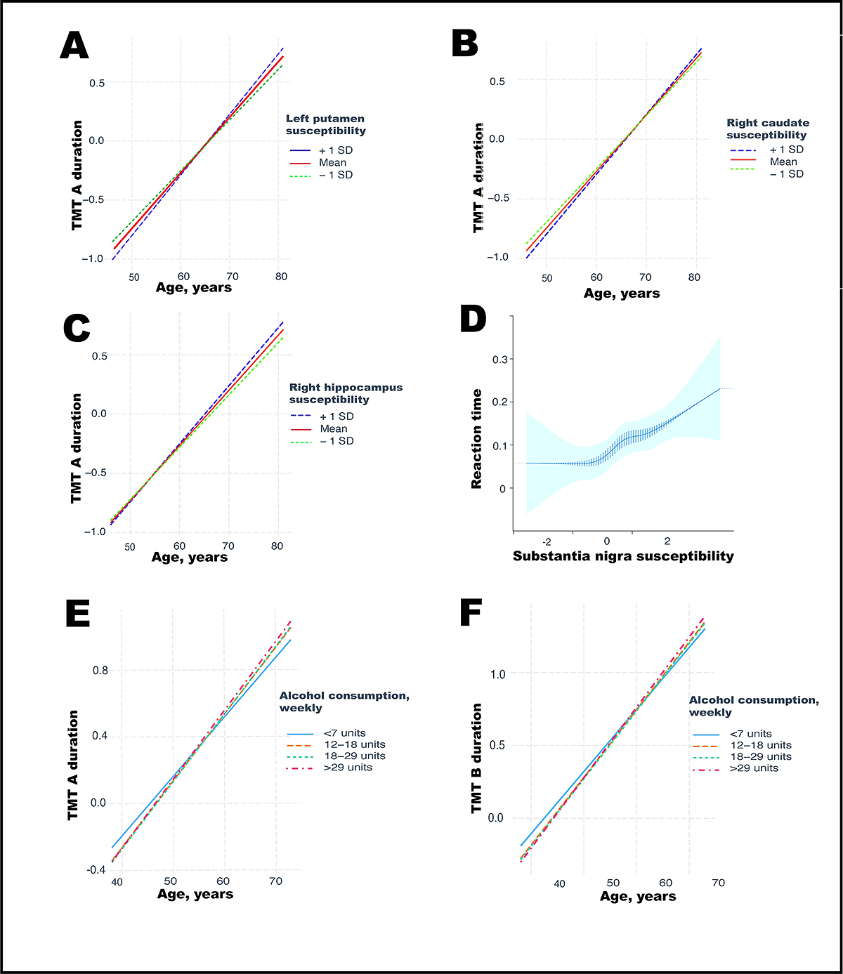
Image source: https://journals.plos.org/plosmedicine/article?id=10.1371/journal.pmed.1004039
Alcohol stimulates the deposition of iron in the brain in two ways:
- The first way — iron immediately accumulates in nerve cells under the influence of alcohol. The contribution of this pathway to the deposition of cerebral iron is 68%. Scientists suggest that alcohol contributes to an increase in the permeability of the blood-brain barrier to iron, as it happens in patients with cerebral autosomal dominant arteriopathy. Iron accumulation in the brain can also contribute to dopamine surges after drinking alcohol.
- The second way is that iron is first deposited in the liver, then the level of iron content in the whole body increases, and then, together with the blood flow, iron enters the brain and accumulates there. The contribution of this pathway to the deposition of cerebral iron is 32%. Scientists note that iron accumulation in the liver is more indicative of alcohol consumption than fatty liver disease or fibrosis (cirrhosis of the liver).
For clarity, scientists schematically depicted these processes:
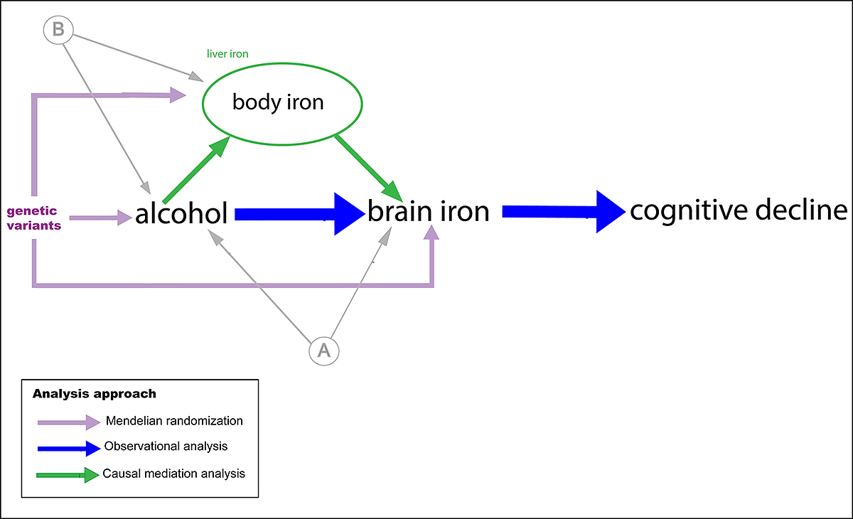
Image source: https://journals.plos.org/plosmedicine/article?id=10.1371/journal.pmed.1004039
High levels of iron in the liver (systemic iron) stimulate iron deposition in the brain. Moreover, every 8 grams of alcohol in ethyl equivalent consumed weekly increases the amount of iron in the liver by 5%.
How Alcohol Promotes Iron Accumulation, Disrupts Iron Balance and Stimulates Cell Death
The Role and Regulation of Iron in The Body
The body needs iron for critical functions:
- in hematopoiesis – for the synthesis of hemoglobin;
- in the life of all types of cells – for DNA synthesis, respiration and energy metabolism, for the production of enzymes;
- in higher nervous activity – to produce neurotransmitters such as dopamine, serotonin, and adrenaline.
However, excess iron is toxic. It causes cell death due to excessive accumulation of lipid peroxides and oxidative stress. The process can cause organ failure.
The biology of our body is such that it stores much-needed iron to recompense its lack. However, there is no physiological way to remove excess iron – only through blood loss. The body loses a significant amount of iron through peeling the epithelium’s stratum corneum.
The amount of iron stored in the body is regulated by the hepatic hormone hepcidin, which controls the ability of intestinal epithelial cells to extract iron from food. Hepcidin changes the shape of the intestinal cell receptor, and the cell cannot capture the iron ion. If no hepcidin exists, the cell successfully captures iron and enters the body. Therefore, when there is an excess of iron in the body, much hepcidin is produced.
Alcohol Disrupts Iron Regulation and Stimulates Cell Death
Alcohol interferes with hepcidin production, thereby disturbing the balance of iron in the body. Therefore, in people who drink, even when the body is overloaded with iron, little hepcidin is produced, and the body continues to accumulate iron.
The toxicity of excess iron in cells was discovered in 2012. American scientists from the University of California found that due to excess iron ions, a lethal dose of lipid reactive oxygen species (ROS) accumulates in cells.
Cells usually use ROS to fight foreign particles: viruses, bacteria, microbes, and cancer cells. However, in this case, excess ROS kills the cell itself as a result of oxidative stress: the integrity of the cell is violated, and the permeability and fluidity of the membrane increase.
This process of programmed cell death, the root cause of an increased iron concentration, has been called ferroptosis.
Thus, one of the ways of the negative impact of alcohol on the brain, thinking and cognitive functions looks like this:
- alcohol suppresses the production of the hormone hepcidin, which regulates the amount of iron in the body;
- as a result, the body accumulates excess iron;
- a significant part of excess iron accumulates in the brain;
- excess iron kills nerve cells as a result of ferroptosis.
Ways to Neutralize Iron Excess
In clinical practice, drugs reduce excess iron in the body – through iron chelators. However, there are many complications and side effects when using them. Therefore, these drugs should only be used as prescribed by a doctor.
Flavonoids, a large class of natural compounds found in plants, can reduce iron overload in the body. Scientists continue investigating flavonoids’ chelating and antioxidant properties to neutralize excess iron. Flavonoids can minimize iron accumulation in tissues and organs and promote its excretion from the body. In addition, flavonoids inhibit active oxygen, scavenge free radicals, and can reduce the concentration of lipid peroxides that lead to cell damage.
Conclusions
The negative effect of alcohol on the brain is significantly higher than previously thought. There is no dose of alcohol that is safe for the brain. All alcoholic beverages are harmful: wine, beer, spirits, and combinations. Under the influence of alcohol, the volume of the brain decreases, and its work worsens.
Alcohol stimulates iron deposition in the brain, characteristic of neurodegenerative diseases such as Alzheimer’s disease, Parkinson’s disease, and dementia. Under the influence of alcohol, the volume of the brain decreases, and the ability for logical thinking, learning, and planning decreases. Also, the reaction time slows down.
Reference
- No safe level of alcohol consumption for brain health: observational cohort study of 25,378 UK Biobank participants
- Moderate alcohol consumption as risk factor for adverse brain outcomes and cognitive decline: longitudinal cohort study
- Associations between moderate alcohol consumption, brain iron, and cognition in UK Biobank participants: Observational and mendelian randomization analyses
- Hepcidin and iron homeostasis
- Is the iron regulatory hormone hepcidin a risk factor for alcoholic liver disease?
- Ferroptosis: An Iron-Dependent Form of Nonapoptotic Cell Death
- Molecular targets and therapeutic interventions for iron induced neurodegeneration
- Role of Flavonoids in the Treatment of Iron Overload