SARS appeared on November 16, 2002, in Foshan, Guangdong province, China. It was spreading to Southeast Asia and then around the world.
Inappropriate personal protective equipment against SARS led to the infection of 80 health workers at Peking University people’s hospital during the initial outbreak in 2003.
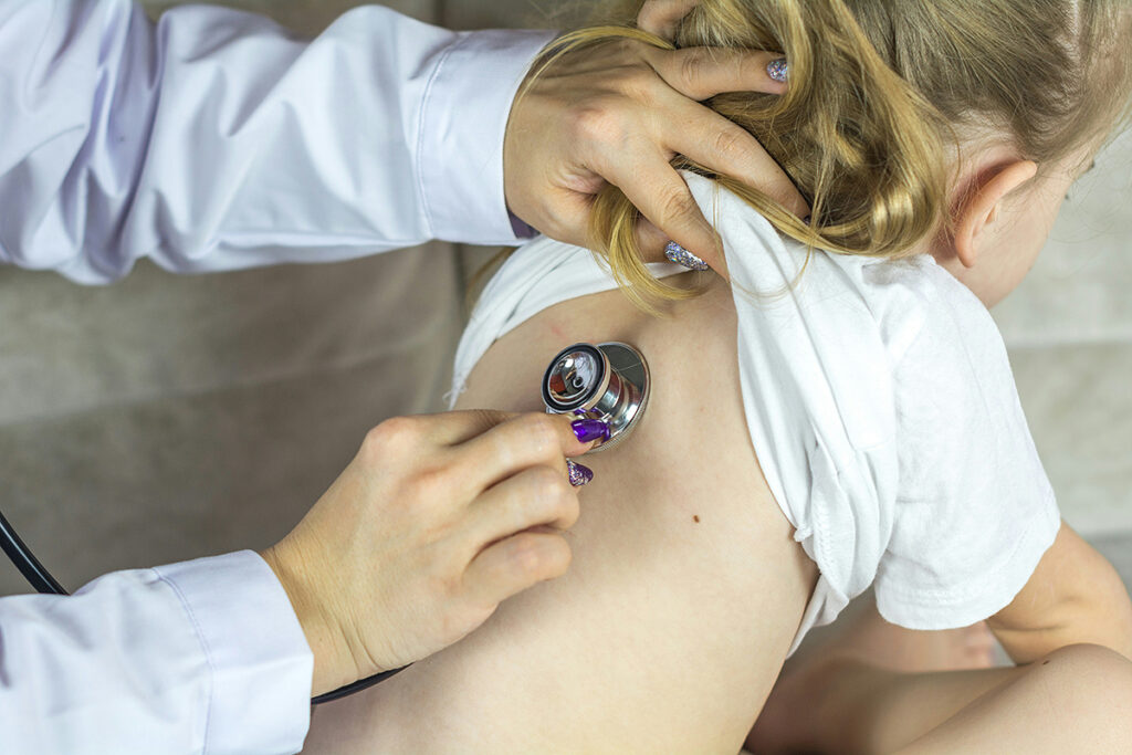
Pneumonia was the main x-ray sign of SARS. Pulse therapy with high doses of anti-inflammatory hormones glucocorticoids was used to treat inflammation. A side effect of therapy was the femoral head necrosis.
Health workers who contracted SARS at Peking University people’s hospitals received the same treatment regimens and rehabilitation plans.
The most severe consequences after rehabilitation from atypical pneumonia were necrosis of the femoral head and pulmonary fibrosis. The 2020 study summarizes the results of a 15-year follow-up of the lung and bone condition of patients who have undergone SARS.
The study involved 71 patients. Patients were hospitalized for 10 to 70 days. As a treatment, methylprednisolone was given intravenously at a daily dosage of up to 800 mg.
Patients underwent annual lung CT and hip MRI scans from 2003 to 2018 together with lung function tests in 2006 and 2018.
Lung recovery was assessed using CT lung scan and functional lung tests. Recovery after necrosis of the femoral head was evaluated using hip MRI and hip function questionnaires.
Functional lung tests
Functional tests were performed to assess the lung condition of patients who had undergone SARS. Among them:
- IVE 25-75% – instantaneous volume expiratory rate of 25-75 % of the FVC, normally > 80%, where the FVC is the forced lung’s vital capacity. It is the difference between the air volume in the lungs at the beginning and end of forced expiration
- FEV1/FVC – the index of the presence or absence of deterioration of airway patency, normally 75-80%, where FEV1 — the volume of forced exhalation for one (first) second.
Lung tests results in 2006:
- Restrictive respiratory failure was detected in 10 of 46 (21.74%) patients. Respiratory failure by restrictive type limiting your maximum depth of inspiration. Restrictive respiratory failure occurs due to lung tissue damage and the inability of the lungs to fully expand during inspiration.
- A decrease in the diffuse lung capacity was found in 16 of 46 (34.78%) patients. Diffuse lung capacity (DL) — the ability of gas to pass from the alveoli to red blood cells. The DL shows how efficient the gas exchange is. A decrease in DL occurs due to the thickening of the alveolar-capillary membrane, a decrease in the alveolar volume involved in gas exchange, and violations of pulmonary blood flow.
Results of lung tests in 2018:
- 1 out of 52 (1.92%) patients had obstructive respiratory failure. Respiratory failure of the obstructive type is characterized by shortness of breath with difficult exhalation. Obstructive respiratory failure occurs due to a bronchi patency violation.
- None of the patients were found to have restrictive respiratory failure.
- The number of patients with impaired IVE values increased by 25%-75%. Violations were found in 16 of 52 (40.38%) patients.
- In 18 (38.46%) patients, there was a decrease in the diffusion capacity of the lungs. Compared to the 2006 test results, the percentage of patients with reduced DL increased slightly, but the difference was not statistically significant.
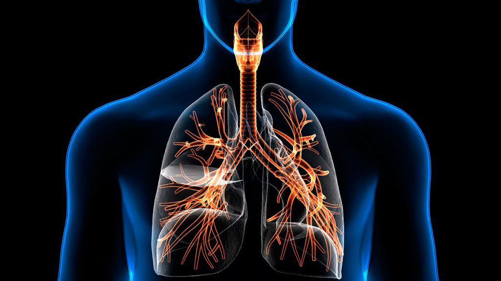
CT of the lungs
CT scans of the lungs of 71 SARS patients showed that:
- 27 patients had a Ground-glass opacity (GGO) symptom for 15 years. GGO-numerous lung tissue seals. GGO is defined on computed tomography as a decrease in the transparency of the lungs with a vascular and bronchial pattern on a matte background of lung tissue. GGO occurs due to the interalveolar partitions thickening.
- In 2003, two patients did not have any abnormalities on CT scans of the lungs, but in 2004 and 2007 there were changes similar to GGO.
- From 2003 to 2018, the percentage of lung tissue lesions gradually decreased.
- Recovery of lung damage occurred mostly between 2003 and 2004 and then remained stable until 2018.
Lung tests and CT scans results
A total of 35 patients underwent lung function tests in both 2006 and 2018. 6 months after infection in 2003, the cohort was divided into two groups: normal CT results and abnormal CT results. The parameters of the pulmonary function test were compared between a group of normal CT results (22 patients) and a group of abnormal CT results (13 patients). Results:
- Over the past few years, no significant changes in lung function were observed in the group of abnormal CT results.
- Between the two groups, only FEV1/FVC and the MOS value of 25% -75% significantly decreased.
- Other parameters, including total lung capacity and carbon monoxide diffusion capacity, did not change significantly 15 years after SARS infection.
- Patients in the normal CT results group had better lung function in 2018 than in 2006.
Tests conclusions
Lungs affected by SARS recover to a greater extent within 1 year after the rehabilitation.
Lung function in 2018 was basically the same as in 2006, with moderately impaired diffusion capacity. Although there was no significant recovery, it is necessary to consider the natural deterioration of lung function over 15 years.
Lung function in patients with normal CT results after recovery in 2003 was significantly better than in patients with abnormalities. This means that lung function can be improved to a greater extent if the acute phase of infectious viral pneumonia is effectively managed.
MRI of the hip
High doses of the anti-inflammatory hormone glucocorticoids were used to treat SARS-induced lung inflammation. A side effect of this therapy was necrosis of the femoral head. Previous studies have shown that subchondral osteonecrosis after systemic steroid treatment was observed in 5-10% of SARS patients and that cumulative steroid dose was a risk factor for osteonecrosis.
To determine the stage of femoral head necrosis, we use the international classification of femoral head osteonecrosis ARCO (5 stages).
Hip MRI in 2003 showed:
- A presence of femoral head necrosis in 15 out of 71 patients.
- In this cohort, 7 patients had unilateral osteonecrosis, then 8 had bilateral osteonecrosis.
- The total number of affected extremities with osteonecrosis is 23 since some patients had bilateral lesions.
MRI results from 2003 to 2018:
- The volume of osteonecrosis:
- rapidly decreased from 2003 (38.83 ± 21.01)% to 2005 (30.38 ± 21.23)%;
- slowly decreased from 2005 to 2013 (28.99 ± 20.59)%;
- increased until 2018 (25.52 ± 15.51)%.
- Among 23 limbs in 15 patients with femoral head necrosis, the osteonecrosis stage was stabilized during follow-up in 16 limbs of 10 patients (3 at stage II A, 8 at stage II B, 4 at stage II C, and 1 at stage III C).
- The condition of one limb has improved from stage IIB to stage IIA.
- In 6 limbs, 4 patients had progressive deterioration.
Evaluation of hip function on the Harris scale
40 patients were assessed for hip function in March 2018:
- In 13 patients with osteonecrosis of the femoral head, the average score was 69.69 ± 13.72.
- In 27 patients without osteonecrosis, the average score was 78.70 ± 11.94.
- Of the 40 patients, 29 (72.5%) had relatively stable hip function during 15 years of follow-up.
- Moderate deterioration was observed in 7 patients (17.5%).
- 4 patients (10%) developed arthritis accompanied by poor mobility.
Conclusions based on MRI results and joint function assessment
The condition of femoral head necrosis caused by high-dose steroid treatment for viral pneumonia can remain stable for a long time.
Necrosis of the femoral head can become reversible.