One of the treatment options for severe patients with COVID-19 coronavirus disease is plasma from convalescents. When searching for potential plasma donors with PCR-confirmed SARS-CoV-2 infection, German scientists found that 17% of the volunteers had borderline or negative results for IgG antibodies to SARS-CoV-2 (antibody ratio <1.1). The scientists decided to observe these volunteers by repeating antibody tests and evaluating T-cell immunity using ELISpot's interferon-gamma assay.
ELISpot’s enzyme-linked immunosorbent assay will help understand how many people may still be infected with the SARS-CoV-2 coronavirus. ELISpot identifies patients with adaptive T-cell immunity, which, like IgG antibodies, can protect against coronavirus infection.
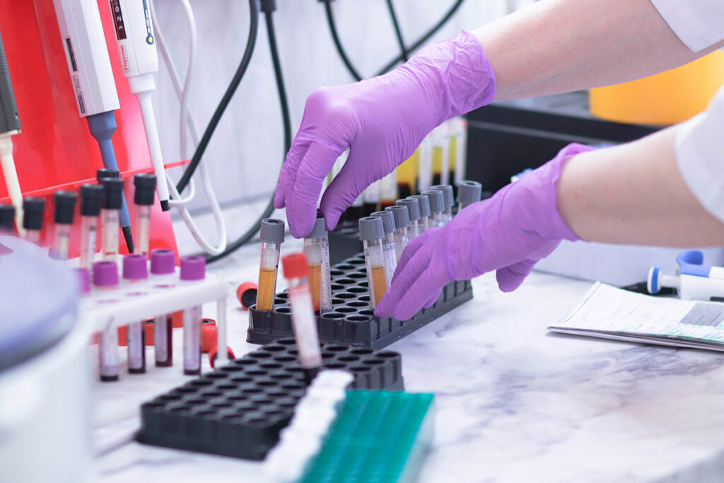
ELISpot measures cellular immunity for 1 day at the level of individual cells, determines functional cells. The results of a study on volunteers with confirmed SARS-CoV-2 infection suggest that most people with undetectable systemic IgG levels may be protected by specific T-cell immunity.
The essence of the IFN-γ ELISpot analysis
T-lymphocytes are the cells responsible for the immune response to an invading pathogen. They recognize and destroy infected cells and stimulate the production of antibodies. When prompted by an antigen, T-lymphocytes produce interferon-gamma (IFN-γ). ELISpot enzyme immunoassay determines the presence of interferon-γ in the blood serum.
SARS-CoV-2 proteins: S1/S2 and membrane protein M were used as antigens to stimulate T-lymphocytes. With the help of the spike protein S, the coronavirus enters the cell. The S protein is the main target of neutralizing antibodies. The S1 fragment of the S protein is responsible for binding the virus to the cell, and the S2 segment is responsible for the fusion of the viral membrane with the cell membrane. S1 is the most specific antigen for the diagnosis of COVID-19. The S2 fragment can cross-react with the S protein of the SARS-CoV-1 and MERS-CoV coronaviruses. However, the infection rate of SARS-CoV-1 and MERS-CoV in Europeans is low. The second stimulus of T-lymphocytes is the membrane protein M. It plays a central role in the assembly of viral particles and is most likely a target for cross-reactive T-cells.
Responses of antibodies and T-lymphocytes in COVID-19 recovering group
Using the SARS-CoV-2-specific ELISpot, the researchers analyzed T-lymphocytes’ responses in a group of 78 potential plasma donors with PCR-confirmed SARS-CoV-2 infection. Negative control – 22 people without symptoms associated with COVID-19, and without household contact with infected patients.
The average interval between the onset of symptoms and the first blood collection in 78 donors was 60 days (range 22-112 days). 28 donors have later tested again, an average of 75 days (24-154 days) after the onset of symptoms. The average interval between the two blood samples was 25 days (range 10-61 days). The ratio of antibodies >1.1 was considered positive, from ≥0.8 to <1.1-borderline, <0.8-negative. The results of the analysis of antibodies:
- 13 of the 78 (17%) donors had borderline or adverse effects (ratio <1.1) for IgG antibodies to SARS-CoV-2.
- Repeated testing in 10 participants with a ratio of <1.1 showed that the majority of antibody results (9/10) remained similar.
- In donors with a higher antibody ratio, the values also remained at the same level.
- In 1 volunteer with a borderline attitude, the value increased over time and became optimistic.
Neutralizing antibodies were not detected in all participants with an antibody ratio of <0.8.
Cellular immunity was determined from 24 to 154 days after the onset of COVID-19 symptoms parallel with antibody testing. The T-lymphocyte response was determined by IFN-γ ELISpot analysis separately for each of the different stimuli: the S1/S2 protein-peptide pools, the M membrane protein, and the S1 protein antigen. Results of the analysis of T-cell immunity:
- None of the ELISpot responses differed significantly between the 9 participants with undetectable antibody responses (ratio <1.1) and the 15 participants with high antibody responses (ratio >3).
- ELISpot responses to all stimuli in donors were significantly higher than in the negative control group.
- The strength of responses to the S1 protein was higher in the group with an antibody ratio >3 than in the group with a rate of <1.1.
- The strength of responses to the S1/S2 peptides was slightly higher in the group with an antibody ratio >3 than in the group with a ratio <1.1, and the M - peptide was similar.
- The strength of responses to the S1/S2 peptides as a whole was higher than to the S1 protein alone.
- The strength of T-cell responses to stimuli in ascending order: S1, S1/S2, M. Such responses were observed both in the group with an antibody ratio >3 and in the group with a ratio <1.1.
- Donors with an antibody ratio of <1.1 showed stable responses directed at the M protein, while patients with an antibody ratio of>3 had durable responses to the S1/S2, S1, and M proteins.
Conclusions
In potential plasma donors with undetectable antibodies against the S1 SARS-CoV-2 protein, the T-cell response to the M membrane protein was more robust than the S1 protein. Perhaps the T-cells of these donors preferentially target peptides involved in the assembly of viral particles rather than binding the virus to the cell. However, this hypothesis needs to be confirmed.
Responses to the S1/S2 peptides were more substantial than the S1 protein, possibly because the S2 antigen macromolecule fragments elicited a more robust immune response.
Interestingly, the differences between participants with an antibody ratio <1.1 and >3 appear to be more pronounced after stimulation with the S1 protein than with the S1/S2 peptides. Thus, in addition to the M protein, the S2 protein may be an additional target for T-cell responses, especially in participants with undetectable T-and B-cell responses against the S1 protein.
Finding that the T-cell response to the S1 protein, which is most specific for SARS-CoV-2, was relatively low raises the question of potential cross-reactivity after stimulation with S1/S2 or M peptides.
Cross-reactivity has previously been observed for SARS-CoV-1 and SARS-CoV-2 antibodies. Similarly, SARS-CoV-2 cross-reactive T-cells may occur due to contact with common coronaviruses. It may affect the specificity of ELISpot tests. However, a recent PCR-confirmed SARS-CoV-2 infection may have resulted in a higher rate of SARS-CoV-2-reactive T-cells in potential plasma donors than in those who had other common coronaviruses. Cross-reactive T-cells can also protect against SARS-CoV-2 infection, especially in children and young people with frequent social contact. A study by Braun et al. It was shown that 34% of healthy plasma donors have pre-existing SARS-CoV-2 S-cross-reactive CD4+ T-cells, and the survey by Grifoni et al. It found cross-reactive T-cells in 40% -60% of individuals who were not exposed to the SARS-CoV-2 coronavirus. However, due to the possibility that cross-reactivity interferes with ELISpot assays, the scientists tested the T-cell responses in the negative control group. The answers were negative.
Cellular immunity to any of the SARS-CoV-2 antigens was found in 7 of the 9 (78%) participants who had an antibody ratio of <1.1. In comparison, 12 of the 15 (80%) participants with an antibody ratio >3 had detectable cellular immunity. Considering all potential plasma donors with PCR-confirmed SARS-CoV-2 infection (also with an antibody ratio of 1.1-3), 22 of the 28 (79%) donors had T-cell immunity.
Thus, T-cell immunity against SARS-CoV-2 was observed in most participants who were PCR-positive for SARS-CoV-2, with undetectable IgG antibodies to the S1 protein. In this group, T-cell immunity was more strongly directed against protein M than against protein S1.
Does T-cell immunity protect against re-infection with coronavirus?
The absence of IgG antibodies to SARS-CoV-2 does not mean that the person did not have a coronavirus. The assessment of cellular immunity can complement the data on the humoral response. Previous studies have shown that the magnitude of the antibody response to SARS-CoV-2 depends on the duration and strength of exposure to the virus. The lack of sustained systemic IgG responses may indicate a mild and temporary SARS-CoV-2 infection that has been virtually eliminated, for example, by the innate immune system. However, it is necessary to determine whether this immune response led to protective immunity from the coronavirus.
Chandrashekar et al.: Almost complete protection was observed in 9 rhesus macaques after SARS-CoV-2 infection. After initial neutralization of the virus during re-infection, the animals decreased the average viral load compared to the primary disease and a stable humoral and cellular immune response. Deng et al.: It was reported that when re-infected with SARS-CoV-2, 4 rhesus macaques experienced a temporary increase in body temperature, but without a viral load. Kirkcaldy et al.: Limited evidence of re-infection among COVID-19 survivors has been reported. A Singapore study found that SARS-CoV-2-specific T-cells were detected in the majority of recovered patients. Data on the previous SARS-CoV-1 coronavirus showed that T-cellular immunity was detected for more than 17 years after infection. Scientists in Sweden and France observed a T-cell response against SARS-CoV-2 in patients without antibodies to the coronavirus.
IFN-γ ELISpot data obtained on average 2 months after the onset of SARS-CoV-2 symptoms shows that 79% of participants had detectable T-cell immunity, which is in good agreement with previous data structurally related SARS-CoV-1 coronavirus. It means that in recovering patients with undetectable IgG to SARS-CoV-2, T-cells can provide immunity.