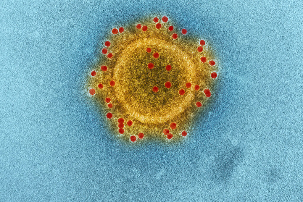
Human coronaviruses were responsible for the epidemics of the last 20 years: severe acute respiratory syndrome SARS-CoV (SARS-CoV) and the Middle East respiratory syndrome MERS-CoV. COVID-19 is a disease caused by the SARS-CoV-2 virus. Many antiviral drugs are being repurposed to treat COVID-19. In this regard, the effectiveness and safety of interferons (IFN) are being studied.
Recombinant and pegylated interferons
Recombinant interferon is an IFN obtained by genetic engineering. Recombinant interferon is safe from infection with blood-borne diseases.
Pegylated interferon is an IFN compound with propylene glycol. Pegylated interferon remains in the body longer.
For the treatment of diseases such as viral hepatitis and multiple sclerosis, recombinant and pegylated IFN-α and IFN-β (interferons type I)are used. The safety and effectiveness of IFN-λ (interferon type III) are also being investigated for the treatment of viral hepatitis.
This review describes the antiviral response caused by IFN in coronavirus infection, as well as innate immune recognition and the ability of SARS-CoV and MERS-CoV to avoid it. The review examines the role of type I and III IFN responses to SARS and MERS, and explores the possibilities and limitations of using interferon for the treatment of COVID-19.
Mechanism of the interferon response
Autocrine signaling: the cell secretes a chemical agent that binds to the receptors of the same cell. It leads to changes in the cell.
Paracrine signaling: signals are transmitted between neighboring cells. Cells produce signaling molecules (paracrine factors) and secrete them into the extracellular environment. Paracrine factors extend over relatively short distances. Factors move to neighbor cells and cause changes in them.
In response to the virus entering the body, cells produce interferon. The innate immune system recognizes viral infections and elicits a reaction from type I (α, β, ε, κ, ω) and type III (λ) interferons. Using autocrine and paracrine signaling, type I interferons bind to their corresponding IFNAR receptor and trigger an antiviral response — the expression of interferon-stimulated genes (ISG). ISGs suppress virus replication. Type III interferons bind to the corresponding IFNLR receptor, which is expressed in epithelial and myeloid cells. The ISG signatures induced by interferon types I and III are similar. ISG expression caused by type I IFN both increases and decreases faster.
Interferons of types I and III make the cell resistant to viruses and trigger an antiviral adaptive immune response.
Some viruses have learned to avoid immune recognition and suppress IFN and ISG functions. Many viral proteins alter the body’s response to interferon. These mechanisms are typical for SARS-CoV and MERS-CoV.
The result of transmitting IFN signals depends on both the virus and the host. For example, type I IFN signals can be harmful due to the systemic Pro-inflammatory effects of interferon. Whether the IFN signal has a protective or pathogenic effect in SARS and MERS depends on the context in which the signal is generated.
Congenital recognition of coronavirus
The innate immune system recognizes pathogens that have entered the body using pattern recognition receptors (PRRS). PRRS recognize pathogen-associated molecular patterns (PAMP) – a set of nucleic acids and virus replication products. These signatures are not present in the original cells of the body.
Two types of PRR recognize viral RNA: Toll-like receptors (TLR) and RIG-I-like receptors (RLR). TLRs are located in the endosome, and RLRs are located in the cytosol. RLR is present in most cells of the body. TLR expression occurs in innate immune cells. Some ISGs (OAS, IFIT) can also recognize and suppress viral RNA.
Genetic studies have shown that susceptibility to coronavirus infection increases if specific PRRS and their signaling pathways are absent. Replication of coronaviruses occurs in the cytoplasm. Therefore, replication products and viral genomes are recognized by the RIG-I and MDA5 cytosolic receptors. Both of these receptors are involved in the detection of the mouse hepatitis virus (mouse coronavirus, MHV). If the RIG-I and MDA5 receptors are absent, interferon induction in response to MHV does not occur. It is possible that these cytosolic receptors also recognize SARS-CoV-2.
TLR receptors: TLR3, TLR7, and TLR8. TLR3 recognizes double-stranded RNA, and TLR7 and TLR8 recognize single-stranded RNA. TLR7 is the most critical receptor that recognizes coronaviruses such as MHV, SARS-CoV, and MERS-CoV. TLR7 is necessary for plasmacytoid dendritic cells to produce IFN-α in these infections. TLR4 expression occurs in innate immune cells. TLR4 recognizes viral docking proteins, in particular, the protein of the respiratory syncytial virus that affects the lower respiratory tract. A study in mice demonstrated that TLR4 deficiency increases morbidity and mortality after infection with MHV and SARS-CoV.
The adapter – a protein that transmits a signal from the receptor to the effector, which regulates the activity of specific proteins.
Adapter-molecules for TLR-MyD88 (for TLR4, TLR7, TLR8) and TRIF (for TLR3, TLR4). Adapter-molecules are an essential part of the defense mechanism against coronavirus infections.
MAVS is a mitochondrial antiviral signaling protein. MAVS is further along the signal path from the RLR.
In a 2008 study, mice were infected with the rMA15 virus (adapted to mice with atypical pneumonia). The study showed that mice with MyD88 deficiency are unable to control virus replication and die. The incidence of TRIF-deficient mice is comparable to that of MyD88-deficient mice.
A 2014 study on mice showed that if MyD88 signals are absent in MERS-CoV, then the decrease in viral load slows down, and pulmonary pathology increases. Tlr7−/− mice have reduced IFN expression compared to wild-type mice. This effect is not observed in Mavs−/− mice. It means that the TLR7 – MyD88 signaling pathway is the main one for innate immune recognition of MERS-CoV.
NF-κΒ is a nuclear factor, a protein complex that controls DNA transcription, cytokine production, and cell survival.
IRF3, IRF7 – interferon regulation factors.
Innate virus recognition triggers two processes:
- production of Pro-inflammatory cytokines (IL-1, IL-6, TNF-α) caused by NF-κΒ;
- the creation of IFN types I and III induced IRF3 and IRF7.
The response of IFN I and IFN III to SARS-CoV-2 is feeble, and ISG expression is limited. It activates chemokines and Pro-inflammatory cytokines.
In addition to the fact that the virus suppresses the IFN response, the level of Pro-inflammatory cytokines depends on age. In the SARS-CoV study on macaques, older macaques had a higher degree of pulmonary pathology and expression of pro-inflammatory cytokines compared to young macaques. Lower expression of type I IFN was also observed. These results are in line with another study showing that older human monocytes have reduced the production of IFN I and IFN III in response to influenza. At the same time, the initial level of Pro-inflammatory cytokines is maintained. In the process of aging, there is an imbalance between the Pro-inflammatory and interferon responses. This fact plays a vital role in the course and outcome of COVID-19.
Coronavirus alters the innate antiviral response
Type I and III interferons cause the expression of antiviral ISGs to make cells virus-resistant. But coronaviruses can avoid immune recognition and suppress the interferon response. That explains the high pathogenicity of coronaviruses.
Coronaviruses can suppress any stage of innate immunity:
- innate recognition;
- the production of interferon;
- interferon signaling pathway;
- antiviral effect of ISG.
IFN and coronavirus infections
Patients with SARS had no interferon response. At the same time, the production of chemokines and cytokines remained stable. In vitro studies confirm that SARS-CoV infection reduces the production of type I IFN. In a 2020 study, blood serum was taken from patients with COVID-19. The result is the same: there are no interferons of types I and III, Pro-inflammatory cytokines and chemokines are elevated.
A 2010 SARS-CoV study has observed that interferon expression lags behind that of Pro-inflammatory cytokines. That is, the response to interferon is not absent but delayed. A study of SARS-CoV in mice showed that several hours passed from the peak of viral load to the expression of type I interferon in the lungs.
At the same time, a 2007 study found that high levels of IFN-α and ISG in patients with SARS are associated with a severe condition. Even after the disease became less acute, patients with hypoxemia maintained high levels of IFNAR1 and interferon-induced chemokines.
In patients with severe MERS, IFN-α levels were also elevated. In patients with COVID-19, high levels of IFN-α and ISG, correlate with severe disease and high viral load. These facts show that in severe infections, the level of interferon increases while maintaining a high viral load.
SARS studies in mice have shown how genetic factors affect the effect of interferon signals. The most popular laboratory mice C57BL/6 and 129 mice with type I, II, or III interferon knockout were used for research.
In mice, 129 SARS develops in a mild form because interferon signals contribute to increased viral clearance.
Stat1– / – mice (with STAT1 deficiency) have no response to IFNα and IFNγ.
BALB/c – immunodeficient mice that lack the thymus and are unable to produce T-cells.
In Stat1– / – 129 mice, viral control is impaired, so SARS is fatal for them, although it occurs with the same severity as in wild-type mice. In Stat1– / – C57BL/6 mice, the incidence of SARS-CoV was higher. And ifnar1– / – C57BL/6 mice had higher viral titers.
For BALB/c mice, SARS-CoV is fatal, since IFN I signals lead to infiltration of inflammatory monocytes and macrophages in the lungs. SARS symptoms in Ifnar1– / – BALB/c mice were moderate, and the survival rate was 100%.
Early IFN inhibits virus replication. In severe SARS in BALB/c mice, IFN I induction is delayed relative to virus replication. The introduction of IFN I before the peak of viral load enhanced viral control and provided antiviral protection. But the introduction of IFN I after the maximum of the viral load did not lead to this result.
Mers studies in mice have shown that the IFN I signal reduces morbidity and mortality in mice. Ifnar1-/- mice had more tissue damage and worse clinical outcomes than wild-type mice. Blocking IFNAR1 increased viral load and mortality.
Unlike SARS-CoV, with MERS-CoV, the IFN I response is not delayed with virus replication. The reason is that interferon acts differently in SARS and MERS.
Early introduction of type I IFN protects against MERS. In mice that were given IFN-β as a precaution, there were no inflammatory processes and weight loss, and the virus clearance was faster. The introduction of IFN-β before the peak of viral load provided antiviral protection. But the induction of IFN I after the maximum of viral load increased inflammatory responses and mortality.
SARS and MERS studies show that IFN-I response time relative to virus replication determines the course and outcome of the disease. Early response or IFN I administration provides antiviral protection. If the IFN I response is delayed, viral control is reduced, leading to inflammation and tissue damage. The virus can suppress the IFN response. IFN expression decreases with age. Introduction of IFN I is useful only in the initial stages of the disease. It is essential for delayed or reduced IFN I expression.
ACE2 is a docking protein for SARS-CoV.
DPP4 is a docking protein for MERS-CoV.
The course and mortality of SARS and MERS differ in humans and animals. The reason is due to various restriction factors and expression of docking proteins. ACE2 expression increases the incidence of SARS-CoV in mice. But the mouse model of SARS flow differs from the human model. Studies of SARS-CoV and MERS-CoV on non-apes have shown that the pathogenesis of these models differs from humans.
People and animals also have different IFN responses. During SARS in mice and non-apes, the IFN response is disrupted or delayed. People don’t have an IFN response.
The ISG profile in humans and animals is also different. Interferon increases ACE2 expression in humans. In mice, the appearance is less.
IFN response and disease outcome depend on host-specific factors. It is crucial to study the effect of IFN during the entire period of COVID-19 disease.
Type I IFN COVID-19 treatment
Type I recombinant interferons were considered for the treatment of SARS, MERS, and COVID-19. IFN I has shown its effectiveness both in vitro and in vivo for the treatment of SARS and MERS.
IFN-β1b reduced viral load and lung damage in monkeys with MERS-CoV. IFN-β in combination with lopinavir-ritonavir improved lung function, although it did not reduce lung damage in mice with MERS-CoV. IFN-a2b, in conjunction with ribavirin, improved viral control and outcome of MERS-CoV in macaques.
Interferon was also effective against SARS-CoV in animal models. Prevention of IFN-α before infection with SARS-CoV reduced virus replication and pulmonary pathology in macaques.
Clinical studies of interferon use in patients with SARS and MERS have shown more different results. IFN-α, as an adjunct to corticosteroids, improved oxygen saturation and accelerated lung recovery in patients with SARS. IFN-α, in combination with ribavirin, increased survival in patients with MERS 14 days after diagnosis but did not affect survival after 28 days. Combination therapy was not effective in the later stages of SARS. The reason for the different results of using IFN among patients is a limited number of participants in retrospective studies, various combinations of drugs, the use of IFN to varying stages of the disease, and concomitant diseases.
Experience with IFN I for the treatment of SARS and MERS is used for COVID-19. In vitro, SARS-CoV-2 is more sensitive to IFN I than SARS-CoV. Pretreatment of IFN-α and IFN-β cells reduces viral titers. That shows the possible effectiveness of IFN I for the prevention and early treatment of SARS-CoV-2.
In China, the treatment of COVID-19 uses nebulized IFN-α in combination with ribavirin. Nebulized IFN-α acts directly on the respiratory tract.
Research on the effectiveness of IFN I for the treatment of COVID-19 continues. It is comparing the efficacy of subcutaneous injections of IFN-β1a in combination with lopinavir/ritonavir and monotherapy with lopinavir/ritonavir, remdesivir, hydroxychloroquine, and monotherapy with nebulized IFN-β1a.
In a retrospective study, 77 patients with COVID-19 in Wuhan were treated with nebulized IFN-α2b, Arbidol, and a combination of both. IFN-A2B significantly reduced the duration of the detection of viral and inflammatory markers. Another study showed the effectiveness and absence of side effects of recombinant IFN-α nasal drops for the prevention of COVID-19. This study was conducted among 2,944 health workers for 28 days. During this time, no cases were detected.
On the other hand, recent studies have shown that the docking protein for SARS-CoV-2 ACE2 is an ISG that is regulated by IFN-α in upper respiratory tract cells. It indicates that IFN can facilitate the virus’s entry into cells. A retrospective study describes the course and outcome of COVID-19 in 446 patients. The results of the study showed an Association of late IFN administration with slower improvement in lung CT. It is possible that during the peak of viral load in moderately ill patients, the positive and harmful effects of IFN compensate for each other. In severely ill patients, the viral load could reach a peak. Therefore, the expression of ACE2 may not pose as much risk as before the height of the viral load.
IFN type III and respiratory infections
Just like type I IFNS, type III IFNS are expressed when pattern recognition receptors recognize PAMP. Through the jak-STAT signaling pathway, interferons launch an antiviral program.
Type I and type III interferons are not redundant, as they are expressed in different cell types, and type I and type III IFN responses differ:
1) IFN-λ binds to the corresponding IFNLR receptor. While IFNAR is expressed everywhere, IFNLR is expressed by epithelial cells of the respiratory, gastrointestinal, and reproductive tracts and some myeloid cells. It allows you to control the virus directly at the point of its penetration.
2) type I and type III ISG Signatures are similar. But IFN III induces a more stable expression of ISG.
3) only IFN I, in contrast to IFN III, stimulates the expression of the transcription factor of the PRO-inflammatory gene IRF1. IFN – λ is the predominant IFN produced by epithelial cells in the early stages of influenza. IFN-λ binds to the IFNLR of epithelial cells and neutrophils, suppressing virus replication, and not causing inflammation.
Animal studies of the role of IFN III in SARS and MERS have shown that in animals infected with MERS-CoV, TLR7-induced IFN-λ production correlates with viral replication kinetics. In mice infected with SARS-CoV, ISG induction was STAT1-dependent and independent of IFNAR. It indicates that a signal can induce ISG via IFNLR.
The IFN-λ response is protective for respiratory tract infections such as influenza and SARS-CoV. Ifnlr1−/− mice cannot control SARS-CoV replication. IFN I and IFN III responses have an additive effect. Ifnar1−/− Ifnlr1−/− knockout mice with IFNAR and IFNLR receptors have a higher viral load than mice with only one knockout receptor. Ifnar1−/− Ifnlr1−/− mice lack IFN I and IFN III signals. That worsens viral clearance. But the severity of the disease in these mice is lower than in Stat1−/− mice. That indicates the contribution of type II IFN to antiviral protection.
The need for IFN-λ is different in the upper and lower respiratory tract. In the lungs IFN-α and IFN -λ mostly duplicate each other. But in the upper respiratory tract, only IFN-λ creates antiviral protection.
In a study on mice, natural flu infection was simulated. A low dose of the flu virus was injected into the upper respiratory tract. IFN-λ was needed to prevent the spread of the flu virus from the upper respiratory tract to the lungs. IFNLR1−/− mice had a higher viral load. These mice were more infectious than wild-type mice or IFNAR1−/− mice. Both IFN-α and IFN-λ were administered intranasally for prevention. Both interferons suppressed influenza replication in the lungs, but only IFN-λ long-term protected the upper respiratory tract and reduced infection.
Since IFN-λ allows you to control the virus at the site of its penetration, has a more prolonged effect, and does not cause inflammation, it is considered as a possible treatment method for COVID-19.
The flu virus was studied in mice. IFN-λ had a curative effect without side inflammatory effects. When administered prophylactically, IFN-λ provided the same level of protection as IFN-α. When co-administered with the flu virus, IFN-λ provided more excellent protection than IFN-β. When applied after the onset of symptoms, IFN-λ2 created antiviral protection, while IFN-α4 worsened the course of the disease due to the expression of Pro-inflammatory cytokines and infiltration of immune cells.
The role of IFN-λ has also been investigated for SARS-CoV-2. IFNLR1-knockout human intestinal epithelial cells control virus replication even worse than ifnar1-knockout cells.
Cells pretreated with both IFN-β and IFN-λ are more resistant to SARS-CoV-2. A study of SARS-CoV-2 in mice showed that pegylated IFN-λ1, used for both treatment and prevention, suppresses virus replication.
Conclusions
The use of IFN I and III is considered an effective treatment for COVID-19. IFN III suppresses virus replication in the lungs and upper respiratory tract and creates stable antiviral protection. IFN I is more potent than IFN III. But since IFN I causes inflammation, it should only be used at an early stage of the disease. In-time application of IFN I accelerates virus clearance and prevents cytokine storm.
Source
- Type I and Type III Interferons – Induction, Signaling, Evasion, and Application to Combat COVID-19
- MyD88 Is Required for Protection from Lethal Infection with a Mouse-Adapted SARS-CoV
- Rapid generation of a mouse model for Middle East respiratory syndrome
- Exacerbated Innate Host Response to SARS-CoV in Aged Non-Human Primates
- Aging impairs both primary and secondary RIG-I signaling for interferon induction in human monocytes
- Toll-Like Receptor 4 Deficiency Increases Disease and Mortality after Mouse Hepatitis Virus Type 1 Infection of Susceptible C3H Mice


