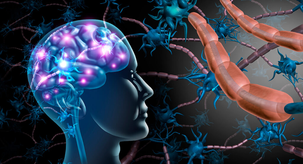
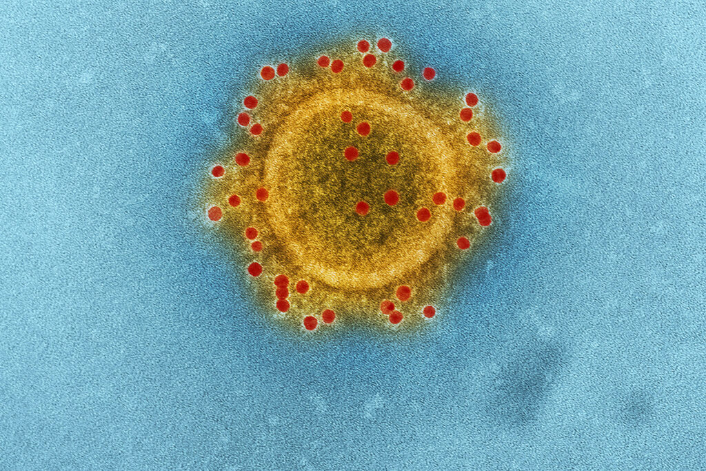
Coronaviruses cause respiratory and intestinal infections in animals and humans. They were not considered highly pathogenic to humans until the outbreak of severe acute respiratory syndrome (SARS) in 2002 and 2003 in China because coronaviruses circulating before that time caused mild infections in humans. Ten years after SARS, another highly pathogenic coronavirus, MERS, has emerged in the Middle East.
Although the SARS-CoV-2 coronavirus has a lower mortality rate (~ 0.7%) than SARS (~ 10%) and MERS (~ 30%), there is increasing evidence that patients hospitalized with the COVID-19 coronavirus disease are affected by the nervous system.
Is the brain vulnerable to the SARS-CoV-2 coronavirus?
To attach to the cell, SARS-CoV-2 uses the ace2 docking receptor. However, for the virus to enter the cell, it is necessary to bind to the ACE2 receptor and process the viral spike protein with the TMPRSS2 protein. In addition to ACE2, SARS-CoV-2 can use Basigin (BSG, CD147) and Neuropilin-1 (NRP1) as docking receptors.
The ACE2 protein has been found in human brain vessels. It was present in the vascular wall’s pericytes and smooth muscle cells and the thalamus’ brain and neurons’ vascular plexus in the middle temporal and posterior cingulate gyrus and hippocampus.
A joint study by American and French scientists has shown that SARS-CoV-2 can infect neurons and cause neuronal death in an ACE2-dependent manner. Another study on brain cells derived from human pluripotent stem cells showed that dopaminergic neurons are particularly susceptible to SARS-CoV-2 infection.
Taken together, these findings show that vascular wall cells can Express ACE2 in the human brain, and SARS-CoV-2 docking receptors are present in several types of brain cells, making the brain vulnerable to the virus.
However, the vasculature of the brain has an antiviral defense system. Besides, the cells of the vessels’ inner surface (endothelium) can perceive the signals of the circulating antiviral protein interferon type I (IFN-I). Both of these mechanisms limit the penetration of SARS-CoV-2 into the brain.
Clinical and pathological studies tested for the virus’s presence in the brain and cerebrospinal fluid gave mixed results. Some studies have found SARS-CoV-2 RNA in brain dissections and cerebrospinal fluid in patients with encephalopathy or encephalitis but at deficient levels. Other studies have failed to detect viral invasion, even though there are signs of inflammation of the cerebrospinal fluid.
Brain-enter pathways for SARS-CoV-2
Olfactory pathway
Loss of smell is a common neurological manifestation of COVID-19. Magnetic resonance imaging (MRI) data from A COVID-19 patient showed an increase in the olfactory cortex’s MRI signal, indicating an olfactory system infection.
It is unclear whether the virus is confined to the olfactory epithelium or reaches olfactory neurons. The virus can enter nerve endings and spread to other brain areas, as described for other coronaviruses. A French study showed that the hypothalamus and its associated regions Express ACE2 and TMPRSS2, needed for SARS-CoV-2 to enter cells. However, in another study, ACE2 and TMPRSS2 were found in the nasal mucosa but were localized to epithelial cells rather than olfactory neurons.
Blood-brain barrier
The blood-brain barrier (BBB) prevents circulating neurotransmitters, toxins, microorganisms, immune proteins, and cells from entering the Central nervous system.
The blood-brain barrier is a standard route for blood-borne viruses to enter the brain. In COVID-19, the virus’s spread into the blood is described, although with a wide frequency range from 1 to 41%. Thus, the virus could enter the brain by crossing the BBB.
Crossing the BBB requires the transport of the virus through the cerebral endothelium, in which the expression of SARS-CoV-2 docking proteins remains unclear. The immunoreactivity of ACE2 was observed in the brain vessels of a patient who died from multiple ischemic infarcts, but the cellular localization was not determined.
The possibility of SARS-CoV-2 penetration through other receptors more widely expressed in the cerebral vasculature, such as NRP1 and BSG, cannot be ruled out. On the other hand, signaling molecules cytokines associated with SARS-CoV-2, including interleukins IL-6, IL-1β, IL-17, and tumor necrosis factor TNF, destroy the BBB and may facilitate viral entry.
Belgian scientists suggest that SARS-CoV-2 causes endothelial infection and inflammation in peripheral vessels, but direct evidence for cerebral endothelial cells has not yet been obtained. The results of the autopsies of patients who died from COVID-19 show rather the absence of bright cerebrovascular inflammation.
Comorbidities are often seen in COVID-19, including cardiovascular or pre-existing neurological diseases, which may increase the BBB’s permeability by themselves or in combination with cytokines. For example, electron microscopy in a patient with COVID-19 and Parkinson’s disease showed viral particles in the frontal lobes’ microvessels and neurons, suggesting that the virus has penetrated through the endothelium. In another patient with Parkinson’s disease, obesity, hypertension, and diabetes, autopsies revealed hypoxic-ischemic neuronal damage, microhemorrhages, white matter damage, and enlarged perivascular space, but no signs of SARS-CoV-2 in the brain.
SARS-CoV-2 can also enter the brain through the median elevation of the hypothalamus and other near-ventricular organs – areas of the brain with a leaky BBB due to holes (Fenestra) in the capillary wall. Although the size of the viral particle (80-120 nm) is larger than the size of endothelial fenestras, preliminary data suggest that the capillaries of the median elevation and tanycytes (cells of the third ventricle of the brain) express ACE2 and TMPRSS, which may contribute to the penetration of the virus into the hypothalamus. Since the hypothalamus is connected to almost the entire Central nervous system, the virus can spread to the whole brain through it.
The infiltration of infected immune cells
Viruses can enter the brain through infected immune cells. Monocytes, neutrophils, and T cells enter the brain through the vasculature, meninges, and vascular plexus. They can be entry points for infected immune cells. Definitive evidence of infection of immune cells with SARS-CoV-2 has not yet been obtained.
Chinese scientists have discovered that SARS-CoV-2 can infect tissue macrophages of the spleen and lymph nodes. Another study showed the presence of viral RNA in macrophages in the bronchoalveolar lavage of patients with COVID-19. However, it remains unclear whether this is due to the virus’s actual reproduction in macrophages or the uptake by phagocytes of virus-infected cells or extracellular virions. Also, studies of the brain of patients who died from COVID-19 did not reveal immune cells’ infiltration.
Thus, SARS-CoV-2 can infect neurons and cause them to die. But the evidence from spinal fluid tests and autopsies does not provide conclusive evidence of the central nervous system’s direct invasion. However, it is impossible to exclude the influence on the median elevation of the hypothalamus and other near-ventricular organs since they can play a role in the disease’s systemic manifestations.
The indirect influence of systemic factors on the brain
COVID-19 affects vital organs, leading to dangerous systemic complications.
Lung damage and respiratory failure
To a greater extent, COVID-19 affects the lungs. It causes alveolar damage, edema, inflammatory cell infiltration, microvascular thrombosis, microvascular injury, and bleeding. SARS-CoV-2 has been detected in epithelial cells of the pulmonary alveoli (pneumocytes) and other epithelial cells.
As a result of lung damage, respiratory failure occurs, which leads to severe hypoxia (acute respiratory distress syndrome). Hypoxia damages the brain. Autopsies of patients who died from COVID-19 showed damage to neurons in the brain regions most vulnerable to hypoxia: the neocortex, hippocampus, and cerebellum.
Systemic inflammation and impaired immune regulation
The critical feature of COVID-19 is an inadequate immune response characterized by hyperactivity of innate immunity with subsequent suppression of immunity (immunosuppression). Improvement in T cell function coincides with remission of symptoms and a decrease in viral load, suggesting a link between immunosuppression and disease severity. Patients with severe disease may develop cytokine release syndrome. These cytokines destroy the BBB. Therefore, viral proteins and molecular complexes from damaged cells can enter the brain from the bloodstream. These molecules will trigger an innate immune response in the brain’s pericytes and resident macrophages. This innate immune response will increase cytokine production and disrupt brain function.
Scientists who have studied pathological inflammation in patients with COVID-19 have shown that although COVID-19 produces a protective IFN type I response, IT can contribute to consciousness changes.
The hypothalamus: the target and the perpetrator of immune dysregulation
The brain, in particular the hypothalamus, can contribute to impaired immune regulation. Some cytokines elevated in COVID-19 (IL-6, IL-1β, and TNF) are potent activators of the hypothalamic-pituitary-adrenal axis (HPA). The hPa axis plays a central role in regulating systemic immune activity and is activated by BBB dysfunction and neurovascular inflammation.
COVID-19 is associated with immunosuppression and lymphopenia. Stroke and brain injuries lead to systemic immunosuppression. Mechanisms of these effects include activation of HPA, which leads to the release of norepinephrine and glucocorticoids. These mediators act synergistically, causing spleen atrophy, T cell death, and a deficiency of natural killer cells (NK cells). Together with the release of calprotectin from damaged lungs, these factors can increase the proliferation of hematopoietic stem cells, which is biased towards myeloid origin cells, leading to lymphopenia and neutrophilia – two key hematological signs of COVID-19. It is important to note that in SARS, HPA activation and glucocorticoid levels correlate with neutrophilia and lymphopenia.
The state of hypercoagulability
Hypercoagulation (blood clot) – is another crucial feature of COVID-19. In a multicenter study, 88% of patients showed signs of hypercoagulation. COVID-19 coagulopathy is characterized by an increased content of D-dimers (fibrin breakdown products that indicate intravascular thrombosis) and increased fibrinogen without significant changes in the number of platelets or an increase in clotting time. Coagulopathy and thrombosis can begin in the lungs and other infected organs with damage to the endothelium, activation of protective blood proteins, increased blood clotting action of IL-6, and the involvement of innate immune cells of neutrophils. Activated neutrophils secrete extracellular traps to fight COVID-19. Extracellular traps are a grid of chromatin and histones that trap cells and platelets in many organs, including the brain, activate clotting and promote intravascular thrombosis.
Systemic organ failure
COVID-19 can damage the kidneys, heart, liver, gastrointestinal tract, and endocrine organs. The resulting systemic metabolic changes, including water and electrolyte imbalances, hormonal dysfunction, and accumulation of toxic metabolites, may also contribute to the effects of COVID-19 on the nervous system. Symptoms of COVID-19: confusion, agitation, headache, etc. Heart damage can affect the brain through a decrease in the cerebral circulation, and it can also cause blockage of blood vessels leading to ischemic strokes.
Neurological manifestations of COVID-19
Numerous neurological disorders have been described in patients with COVID-19. They affect the Central and peripheral nervous system and can occur in patients with severe and asymptomatic SARS-CoV-2 infection. Neurological disorders were present in 30% of patients with COVID-19 who required hospitalization, 45% of patients with severe respiratory diseases, and 85% of patients with acute respiratory distress syndrome (ARDS). In patients with mild coronavirus infection, neurological symptoms are limited to malaise, dizziness, headache, loss of smell and taste. Often, these same symptoms are present in respiratory viral infections, such as the flu. The most severe neurological complications occur in patients in critical condition and are associated with significantly higher mortality.
Encephalopathy and encephalitis
Encephalopathy – is a change in mental status: confusion, disorientation, agitation, and drowsiness. Changes in mental status are rare (<5%), even in COVID-19 patients requiring hospitalization for respiratory illness, but affect most critically ill COVID-19 patients with ARDS.
The critical question is whether the change in mental status represents encephalopathy caused by a systemic disease or encephalitis caused directly by the SARS-CoV-2 virus.
Several cases of COVID-19 infection have been reported that meet the diagnostic criteria for infectious encephalitis: altered mental status, fever, seizures, increased white blood cell count in the cerebrospinal fluid, and focal brain abnormalities during neuroimaging. In two reported cases, the SARS-CoV-2 coronavirus was detected in the cerebrospinal fluid, although only a small amount of viral RNA was detected. In one case of COVID-19, the diagnosis of temporal encephalitis was confirmed by a biopsy that showed perivascular lymphocytic infiltrates and hypoxic damage to neurons, but the presence of SARS-CoV-2 or other viruses in the brain or cerebrospinal fluid was not reported. Most cerebrospinal fluid samples from patients with neurological abnormalities associated with COVID-19 showed no evidence of SARS-CoV-2, and most brain tissue samples taken at autopsy showed no signs of encephalitis.
In addition to encephalitis, most patients with COVID-19 have other causes of changes in mental status. Delirium, States of confusion, and coma are most often found in critical conditions associated with COVID-19, often accompanied by hypoxia, hypotension, renal failure, the need to take large doses of sedatives, and prolonged immobility, and isolation.
Taken together, these findings suggest that SARS-CoV-2 invasion of the brain is a possible but rare cause of encephalopathy.
Ischemic stroke
Stroke rates range from 1 to 3% among hospitalized patients with COVID-19 and up to 6% among patients in critical condition. That is 7 times higher than in patients hospitalized with the flu, even after adjusting for the disease’s severity.
Infections generally increase the risk of stroke. In early reports, strokes in COVID-19 were described in young people, but patients were older in subsequent cases and had numerous concomitant vascular diseases. Thus, it remains unclear whether these strokes were caused by the SARS-CoV-2 coronavirus or occurred in stroke-prone infected people at risk.
Hypercoagulation associated with COVID-19 increases susceptibility to cerebrovascular events. A series of autopsies revealed widespread microthrombi and infarct spots in brain samples. Patients with COVID-19 may be at risk of cardioembolic stroke. Acute cardiac injury and clinically significant arrhythmias were reported in approximately 10% of hospitalized patients with COVID-19 and 20-40% of those who needed intensive care. In rare cases, the SARS-CoV-2 coronavirus can cause myocarditis and heart failure even without significant lung damage. Myocardial damage and arrhythmias in severe infection conditions can lead to heart embolism and brain infarction.
A significant proportion of critically ill patients with COVID-19 may also develop secondary bacteremia and the primary viral disease. In one series of cases, approximately 10% of the patients who required a ventilator had bacteremia. It increases the risk of stroke by more than 20 times. Septic embolism of the brain often leads to bleeding. Post-mortem magnetic resonance imaging of the brain showed signs of bleeding in 10% of cases.
These clinical findings suggest that SARS-CoV-2 can negatively affect the brain through several pathophysiological pathways that lead to damage to brain vessels.
Post-infectious neurological complications
SARS-CoV-2 causes an inadequate systemic immune response, which can have a delayed effect on the nervous system. Manifestations of coronavirus affect both the Central and peripheral nervous system and usually occur after the acute phase of infection subsides.
In the central nervous system, the reported manifestations of COVID-19 resemble classic post-infectious inflammatory conditions, such as acute disseminated encephalomyelitis and acute necrotic hemorrhagic encephalopathy. Several cases of Guillain-Barre syndrome, an immune attack on peripheral nerves, have been reported. The SARS-CoV-2 coronavirus was not detected in any cerebrospinal fluid samples, indicating an immune mechanism rather than a direct infection.
Neurological manifestations associated with intensive care
Changes in mental status in hospitalized patients with COVID-19 are more common in severe illness. Most critically ill patients with COVID-19 require a ventilator, and an agitated state of confusion (delirium) occurs in more than 80% of patients on a ventilator. Patients with ARDS are at an exceptionally high risk of delirium due to hypoxemia, taking high doses of sedatives and paralytics.
Comparison with other viral respiratory infections
Reported post-infectious inflammatory conditions of the nervous system associated with COVID-19 are also observed in other viral respiratory diseases, including coronaviruses. Sometimes with the flu, encephalopathy or encephalitis occurs with signs of the virus’s presence in the cerebrospinal fluid.
Since there are few reported cases of encephalitis in COVID-19 coronavirus infection, SARS-CoV-2 seems more similar to other common respiratory viruses than neurotropic pathogens that target the brain, such as herpes simplex virus.
However, the incidence of thrombotic complications in COVID-19 is much higher than in influenza. In a French multicenter study, patients with COVID-19 and acute respiratory distress syndrome were twice as likely to have thrombotic complications than a matched cohort with ARDS from other causes.
Long-term neurological and neuropsychiatric consequences of COVID-19
Respiratory viral infections are associated with neurological and psychiatric consequences, including parkinsonism, dementia, depression, post-traumatic stress disorder, and anxiety. They do not require brain infection to occur. Inflammation and increased cytokines in sepsis survivors are associated with subsequent hippocampal atrophy and cognitive impairment. In COVID-19, the NLPR3 inflammasome, a protein complex that triggers an inflammatory response, can be activated. Experimental studies suggest a link between inflammasome activation and Alzheimer’s disease. ARDS survivors also often experience long-term depression, anxiety, and cognitive impairment.
Conclusions
The neurological manifestations of COVID-19 are a severe public health concern, not only because of the acute effects on the brain but also because of the long-term harm to brain health. These delayed manifestations are expected to be significant, as they may also affect patients who did not show neurological symptoms in the acute phase.
There are no standard protocols for the treatment of neurological manifestations. Therapeutic efforts should focus on antiviral agents and minimize respiratory and organ failure, hypercoagulation, and impaired immune regulation.
Source
Effects of COVID-19 on the Nervous System


