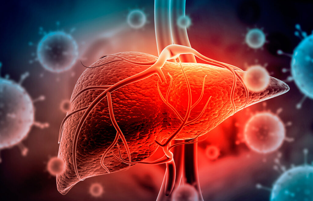
The liver filters the blood, removing or neutralizing dangerous components: bacteria, toxins, carcinogens. Viral and bacterial infections, toxic substances, and injuries kill hepatocytes, which perform filtering functions.
However, the liver is a self-healing organ. Compensatory regeneration of the liver occurs after injuries – the multiplication of hepatocytes. In this way, the size of the liver is maintained in proportion to the size of the body to support the vital functions and homeostasis of the whole organism.
Repetitive liver damage and subsequent compensatory regeneration contribute to the development of cancer. Hepatocellular carcinoma is the most common type of liver cancer.
Taiwanese scientists investigated the effect of type I interferon (IFN-I) on liver regeneration, hepatocyte proliferation, and hepatocellular carcinoma development.
Experts analyzed the reactions to various liver injuries in two groups of mice:
- The first group of mice is standard wild-type mice.
- The second group of mice is genetically modified mice. The cells of these mice are only capable of producing IFN-I. But cells cannot respond to this interferon – they lack the required cellular receptor. Thus, in the genetically modified mice of the second group, the part of the innate immunity responsible for signal transduction of the interferon system is impaired.
Research Results
Scientists have found that in liver injuries when there is no viral infection, the background level of type I interferon can contribute to the development of hepatocellular carcinoma. The mechanism is based on the ability of IFN-I to stimulate the compensatory multiplication of hepatocytes and regulate lipid and glucose metabolism.
Type I Interferon Enhances the Development of Liver Cancer
Experts injected mice with the carcinogen diethylnitrosamine, which causes hepatocellular carcinoma from a single injection. This substance causes mild liver damage and subsequent compensatory regeneration.
As a result, each wild-type mouse developed about 20 tumors in the liver. To increase the production of type I interferon and its effect on cancer, scientists using an adenoviral vector injected mice with the hepatitis C virus protein. As a result, the development of hepatocellular carcinoma in four-week-old wild-type mice accelerated.
In genetically modified mice lacking the type I interferon receptor, the situation was noticeably better. Tumors developed in 70% of the mice. The average number of tumors in one mouse was about 6. The tumors themselves were smaller than in wild-type mice.
Microscopic analysis of the liver, which has a tumor, revealed more lipid vacuoles in wild-type mice than genetically modified mice.
The result of the experiment is shown in the figure:

A – experimental design: two-week-old mice were injected with diethylnitrosamine (DEN), and at the age of 8 months, the state of the liver was studied.
B – the percentage of mice with tumors, number of tumors, size of tumors. WT – wild type mice, Ifnar -/- – are genetically modified mice.
C – photographs of the livers of mice affected by tumors. The scale is 5 mm.
D – stained sections of liver tissue with tumors, scale – 100 μm. The graph shows the average number of lipid vacuoles.
E – immunostaining of tissue sections to determine the activity of the FOXO protein, which regulates the metabolism of lipids, glucose, the cell cycle, and programmed cell death. IFN-I indirectly inhibits FOXO activity. The graph shows that in tumor nodules of genetically modified mice, FOXO activity is higher than in wild-type mice. The scale is 50 microns.
The researchers note that these results indicate that suppression of type I interferon signaling slows down the development of hepatocellular carcinoma.
Blocking Type I Interferon Signals Delays Hepatocyte Reproduction
To assess the role of IFN-I in liver regeneration, scientists removed two-thirds of the liver from experimental mice and observed the process of its recovery. Typically, hepatocyte multiplication peaks occur between 36 and 48 hours after liver resection, and the entire organ is restored on the 7th day after surgery.
Experts found that in wild-type mice, hepatocytes actively proliferated 48 hours after resection. On day 7 postoperatively, the livers of these mice had nearly completed recovery, except for a few cells that continued to divide.
The opposite picture was observed in genetically modified mice with impaired IFN-I signaling. 48 hours after the operation, the multiplication of hepatocytes was much slower than in wild-type mice. On day 7, the livers of the genetically modified mice were still growing, as they had not reached the required size for the body.
Scientists note that these data show that signaling type I interferon stimulates liver regeneration processes.
Type I Interferon Improves Lipid Metabolism and Accumulation in The Liver During Regeneration
After liver resection, changes in lipid metabolism occur to initiate the regeneration process. There is a short-term accumulation of lipid droplets during the recovery process and an increase in glucose levels. Type I interferon regulates these metabolic changes. The body possibly uses the accumulation of lipid droplets as an energy resource for liver regeneration.
To clarify the role of IFN-I in liver repair, the researchers analyzed the presence of lipid droplets in resected mice.
In wild-type mice, a large number of large lipid droplets were observed 24 hours after surgery. However, the genetically modified mice only had tiny lipid droplets. In figure F (scale 20 μm), lipid droplets are colored, and the difference between wild-type and genetically modified mice, in which the signaling of type I interferon is impaired, is visible:

The scientists then analyzed the activity of the membrane protein ADRP, which stabilizes the structure of lipid droplets. Experts found that 24 hours after liver resection, the ADRP activity in wild-type mice was significantly higher than in genetically modified mice.
Scientists suggest that type I interferon signaling is involved in the regulation of lipid metabolism during liver regeneration.
Type I Interferon Regulates Glucose Homeostasis During Liver Repair
After liver resection, changes in glucose metabolism occur. A short-term low level of glucose and the accumulation of lipids initiates the process of organ regeneration.
After two-thirds of the liver was removed, the wild-type mice exhibited decreased blood glucose (hypoglycemia). Within 24 hours after surgery, the glucose level returned to normal. On an empty stomach, the mean blood glucose level was 110 mg / dL.
In genetically modified mice, the mean post-resection glucose level was significantly higher – 140 mg / dL. Moreover, within 24 hours after the operation, it decreased by about 2 times and remained low.
The researchers then investigated the effect of insulin on accelerating liver recovery in genetically modified mice. 2 hours after the operation, specialists injected mice with insulin in an amount of 0.75 U/kg. After that, glucose kinetics in genetically modified mice that received insulin became the same as wild-type mice. After the injection of insulin, the number of multiplying hepatocytes also increased significantly.
Continuing to investigate the factors affecting glucose levels after liver resection, the scientists injected genetically modified mice with a lipid supplement of 20% intralipid emulsion (soybean oil, 100 mg/kg). The supplement increased blood glucose levels and enhanced the liver regeneration process.
The figure shows the results of this experiment:

A – fasting glucose level in wild type (WT) and genetically modified mice with impaired type I interferon signaling (Ifnar -/-).
B – blood glucose level after liver resection.
C – liver sections showing multiplying hepatocytes, scale – 20 μm. The graph shows the levels of hepatocytes in different types of mice.
D – glucose level in genetically modified mice after lipid supplementation (red line).
E – Liver sections showing that genetically modified mice have enhanced liver regeneration after lipid supplementation. Scale – 20 microns.
Scientists note that short-term hypoglycemia can stimulate the accumulation of lipid droplets in the liver to maintain glucose homeostasis. Also, experts suggest that type I interferon signaling regulates glucose homeostasis for timely liver regeneration.
Conclusions
Type I interferon plays an essential role in the multiplication of hepatocytes and liver regeneration in trauma. IFN-I signaling contributes to the timely recovery of the liver, regulates the metabolism of lipids and glucose. However, IFN-I also contributes to the development of liver cancer – a hepatocellular carcinoma. For scientists, the mechanism of the relationship between short-term hypoglycemia, accumulation of lipid droplets, and liver regeneration after injury remains unclear.